Endoscopy Division, Department of Gastroenterology, National Cancer Center Hospital East, Chiba, Japan.
Cancer Sci. 2014 Jul;105(7):857-61. doi: 10.1111/cas.12440. Epub 2014 Jun 3.
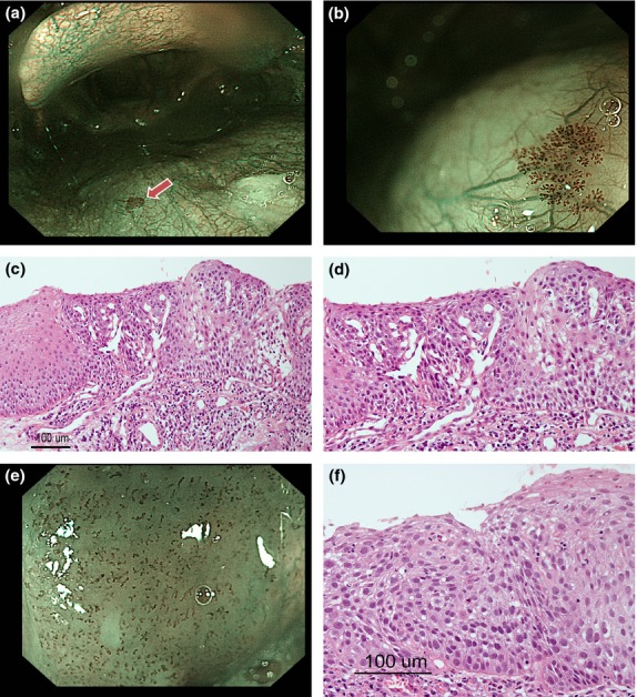
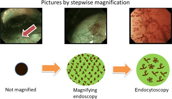
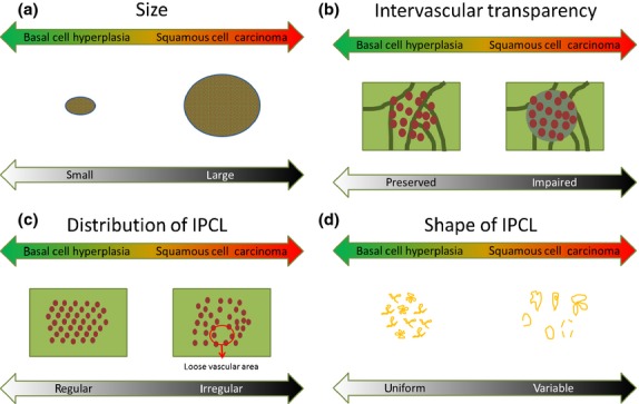
Narrow-band imaging (NBI) has been reported to be useful for detecting superficial-type esophageal or head and neck squamous cell carcinoma (SCC), and in the present study we have used NBI to detect non-carcinomatous lesions, such as basal cell hyperplasia (BCH) accompanied by microvascular irregularities; these non-carcinomatous lesions were pathologically discriminated from squamous cell carcinoma of the pharynx. The aim of the present study was to clarify the endoscopic characteristics of BCH that contribute to the discrimination of superficial-type head and neck SCC (HNSCC). We examined the key endoscopic findings capable of distinguishing BCH from SCC using 26 BCH and 37 superficial-type SCC of the pharynx that had been pathologically diagnosed at our institution between January 2008 and July 2012. The clinicopathological factors were also compared. The size of the BCH lesions was significantly smaller (P < 0.001), and their intervascular transparency was more clearly observed (P < 0.001). Intra-epithelial papillary capillary loop (IPCL) shapes were less variable and monotonous (P < 0.001), and the distribution of the IPCL was more regular with an interval comparable to that of SCC (P < 0.001), although no significant differences in the sharpness of the lesion border, dilatation of IPCL and tortuosity of the IPCL were seen between the BCH and SCC lesions. This study revealed that BCH was an independent entity in terms of not only pathological findings, but also endoscopic findings observed using NBI, such as the regular distribution of IPCL and the preserved intervascular transparency.
窄带成像(NBI)已被报道可用于检测食管浅表型或头颈部鳞状细胞癌(SCC),本研究中我们使用 NBI 检测非癌性病变,如伴有微血管不规则的基底细胞增生(BCH);这些非癌性病变在病理学上与咽鳞癌区分开来。本研究旨在阐明有助于区分头颈部浅表型 SCC(HNSCC)的 BCH 的内镜特征。我们检查了 26 例 BCH 和 37 例我院在 2008 年 1 月至 2012 年 7 月期间病理诊断为浅表型咽 SCC 的患者,以确定能够区分 BCH 和 SCC 的关键内镜表现。还比较了临床病理因素。BCH 病变的大小明显较小(P<0.001),血管间透明度更清晰(P<0.001)。上皮内乳头毛细血管袢(IPCL)的形状变化较小且单调(P<0.001),并且 IPCL 的分布更规则,间隔与 SCC 相当(P<0.001),尽管在病变边界的锐利度、IPCL 的扩张和 IPCL 的迂曲方面,BCH 和 SCC 病变之间没有明显差异。本研究表明,BCH 不仅在病理学发现方面,而且在 NBI 观察到的内镜发现方面,如 IPCL 的规则分布和血管间透明度的保留,都是一个独立的实体。