Woo Sungmin, Kim Sang Youn, Cho Jeong Yeon, Kim Seung Hyup
Department of Radiology, Seoul National University Hospital, Seoul 110-744, Korea.
Department of Radiology, Seoul National University Hospital, Seoul 110-744, Korea. ; Institute of Radiation Medicine and Kidney Research Institute, Seoul National University Medical Research Center, Seoul 110-744, Korea.
Korean J Radiol. 2014 May-Jun;15(3):346-55. doi: 10.3348/kjr.2014.15.3.346. Epub 2014 Apr 29.
To assess the diagnostic value of shear wave elastography (SWE) for prostate cancer detection.
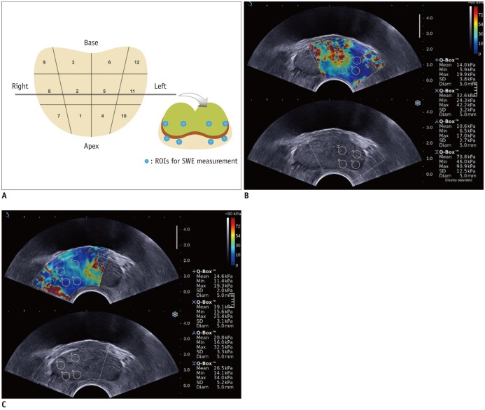
In this retrospective study, 87 patients with the suspicion of prostate cancer (prostate-specific antigen > 4 ng/mL and abnormal digital rectal examination) underwent a protocol-based systematic 12-core biopsy followed by targeted biopsy at hypoechoic areas on grey-scale ultrasound. Prior to biopsy, SWE was performed by placing two circular 5 mm-sized regions of interest (ROIs) along the estimated biopsy tract in each sector and one ROI for hypoechoic lesions. SWE parameters, S (mean stiffness) and R (mean stiffness ratio), were calculated and compared regarding different histopathologic tissues and their accuracy for diagnosing prostate cancer was analyzed. SWE parameters were correlated with Gleason score and were compared between indolent (< 8) and aggressive (≥ 8) tissues in prostate cancer patients.
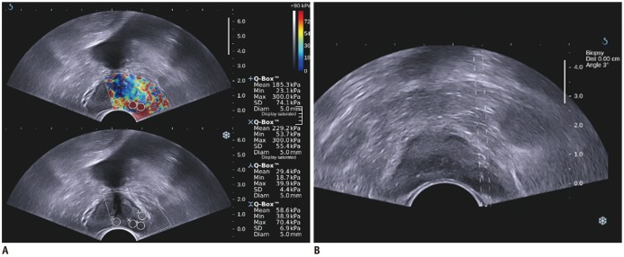
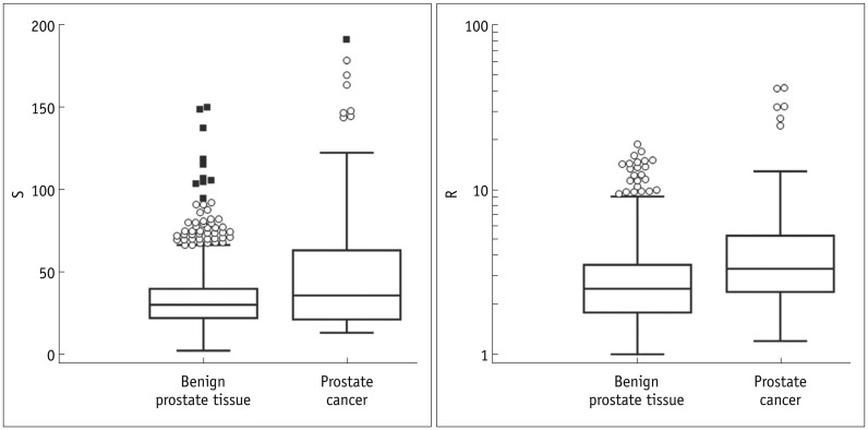
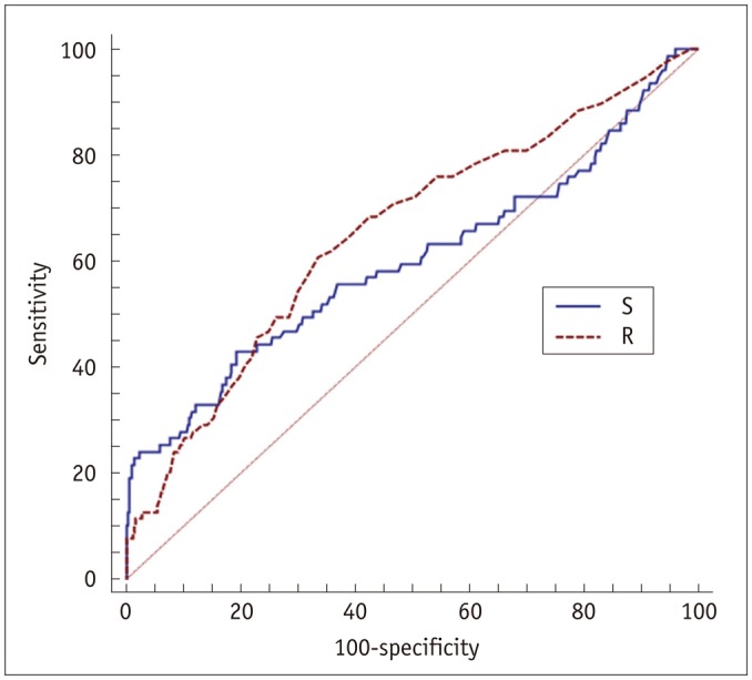
Prostate cancer was detected in 7.5% of 1058 cores in 29.9% of 87 patients. Seven (43.8%) of 16 hypoechoic lesions were confirmed as prostate cancer. SWE parameters were significantly different among the histopathologic entities (p < 0.001). Prostate cancer was stiffer than benign tissues (p ≤ 0.003). Sensitivity, specificity and receiver operating characteristic curve area for diagnosing cancer were 43%, 80.8%, and 0.599, respectively, for a cutoff of S > 43.9 kPa and 60.8%, 66.4%, and 0.653, respectively, for R > 3. Both, S and R showed a significant correlation with Gleason score (r ≥ 0.296, p ≤ 0.008) and were significantly different between indolent and aggressive prostate cancer (p ≤ 0.006).
Shear wave elastographic parameters are significantly different between prostate cancer and benign prostate tissue and correlate with Gleason score.
评估剪切波弹性成像(SWE)在前列腺癌检测中的诊断价值。
在这项回顾性研究中,87例疑似前列腺癌患者(前列腺特异性抗原>4 ng/mL且直肠指检异常)先接受基于方案的系统性12针穿刺活检,随后在灰阶超声下的低回声区域进行靶向活检。活检前,通过在每个扇形区域沿估计的活检路径放置两个5 mm大小的圆形感兴趣区(ROI)以及一个针对低回声病变的ROI来进行SWE检查。计算SWE参数S(平均硬度)和R(平均硬度比),并针对不同组织病理学组织进行比较,分析其诊断前列腺癌的准确性。SWE参数与Gleason评分相关,并在前列腺癌患者的惰性(<8)和侵袭性(≥8)组织之间进行比较。
87例患者中,29.9%的患者在1058针穿刺活检中有7.5%检测出前列腺癌。16个低回声病变中有7个(43.8%)被确诊为前列腺癌。SWE参数在组织病理学实体之间存在显著差异(p<0.001)。前列腺癌比良性组织更硬(p≤0.003)。对于S>43.9 kPa的临界值,诊断癌症的敏感性、特异性和受试者操作特征曲线面积分别为43%、80.8%和0.599;对于R>3,分别为60.8%、66.4%和0.653。S和R均与Gleason评分显著相关(r≥0.296,p≤0.008),并且在惰性和侵袭性前列腺癌之间存在显著差异(p≤0.006)。
前列腺癌与良性前列腺组织之间的剪切波弹性成像参数存在显著差异,且与Gleason评分相关。