Department of Anthropology, University of California San Diego La Jolla, CA, USA ; Psychiatry and Behavioral Sciences, MIND Institute, Department of Psychiatry and Behavioral Sciences, University of California Davis Sacramento, CA, USA.
Department of Anthropology, University of California San Diego La Jolla, CA, USA.
Front Hum Neurosci. 2014 May 20;8:277. doi: 10.3389/fnhum.2014.00277. eCollection 2014.
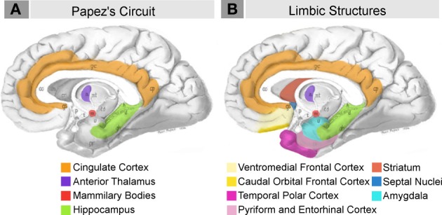
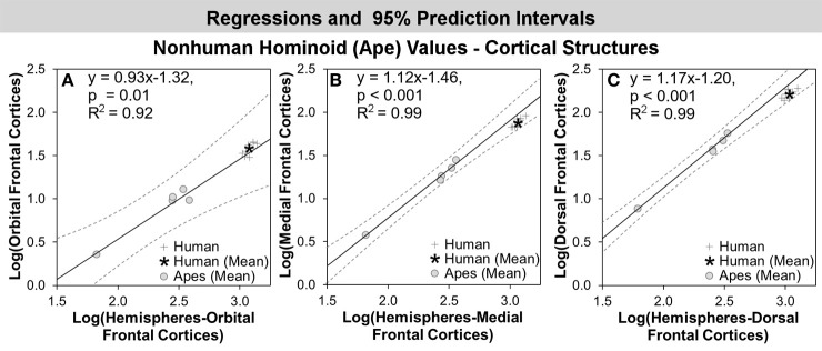
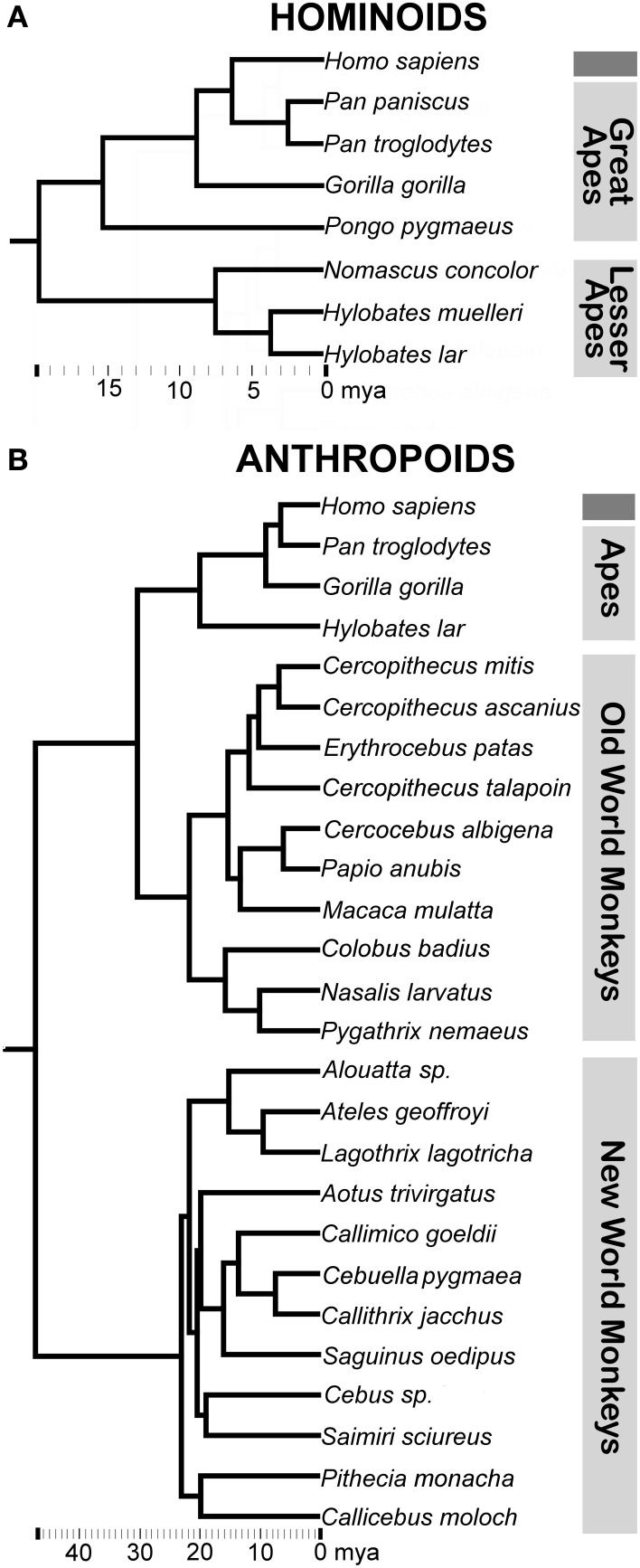
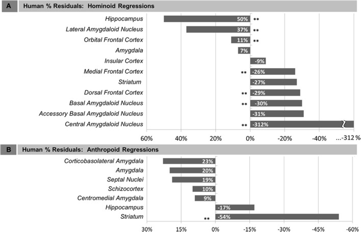
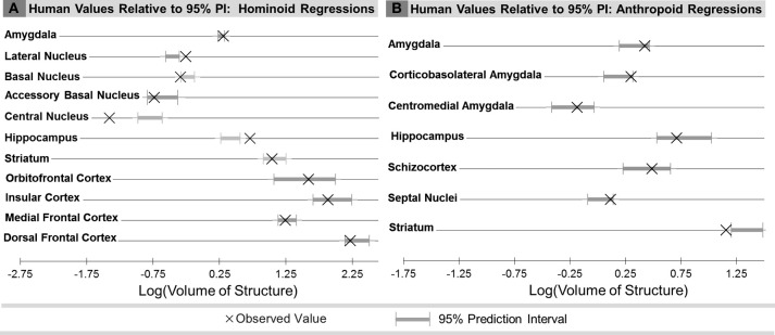
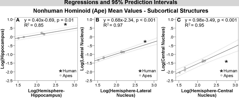
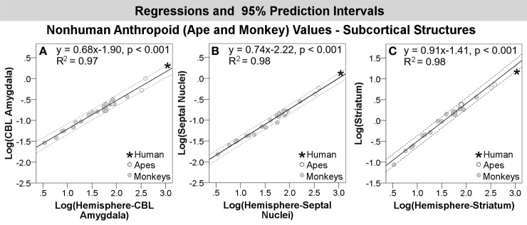
Increasingly, functional and evolutionary research has highlighted the important contribution emotion processing makes to complex human social cognition. As such, it may be asked whether neural structures involved in emotion processing, commonly referred to as limbic structures, have been impacted in human brain evolution. To address this question, we performed an extensive evolutionary analysis of multiple limbic structures using modern phylogenetic tools. For this analysis, we combined new volumetric data for the hominoid (human and ape) amygdala and 4 amygdaloid nuclei, hippocampus, and striatum, collected using stereological methods in complete histological series, with previously published datasets on the amygdala, orbital and medial frontal cortex, and insula, as well as a non-limbic structure, the dorsal frontal cortex, for contrast. We performed a parallel analysis using large published datasets including many anthropoid species (human, ape, and monkey), but fewer hominoids, for the amygdala and 2 amygdaloid subdivisions, hippocampus, schizocortex, striatum, and septal nuclei. To address evolutionary change, we compared observed human values to values predicted from regressions run through (a) non-human hominoids and (b) non-human anthropoids, assessing phylogenetic influence using phylogenetic generalized least squares regression. Compared with other hominoids, the volumes of the hippocampus, the lateral nucleus of the amygdala, and the orbital frontal cortex were, respectively, 50, 37, and 11% greater in humans than predicted for an ape of human hemisphere volume, while the medial and dorsal frontal cortex were, respectively, 26 and 29% significantly smaller. Compared with other anthropoids, only human values for the striatum fell significantly below predicted values. Overall, the data present support for the idea that regions involved in emotion processing are not necessarily conserved or regressive, but may even be enhanced in recent human evolution.
越来越多的功能和进化研究强调了情绪处理对复杂人类社会认知的重要贡献。因此,人们可能会问,参与情绪处理的神经结构,通常称为边缘结构,是否在人类大脑进化中受到了影响。为了回答这个问题,我们使用现代系统发育工具对多种边缘结构进行了广泛的进化分析。在这项分析中,我们结合了使用立体学方法在完整组织学系列中收集的灵长类(人类和猿)杏仁核和 4 个杏仁核核、海马体和纹状体的新容积数据,以及以前发表的关于杏仁核、眶额和内侧前额皮质以及脑岛的数据,以及一个非边缘结构,背侧前额皮质,作为对比。我们使用大型已发表数据集进行了平行分析,这些数据集包括许多类人猿物种(人类、猿和猴子),但灵长类动物较少,用于分析杏仁核和 2 个杏仁核细分、海马体、大脑裂、纹状体和隔核。为了研究进化变化,我们将观察到的人类值与通过(a)非人类灵长类动物和(b)非人类类人猿进行回归运行预测的值进行了比较,使用系统发育广义最小二乘回归评估了系统发育影响。与其他灵长类动物相比,人类的海马体、杏仁核外侧核和眶额皮质的体积分别比人类半球体积的猿类预测值大 50%、37%和 11%,而内侧和背侧前额皮质的体积则分别小 26%和 29%。与其他类人猿相比,只有人类纹状体的值明显低于预测值。总的来说,这些数据支持了这样一种观点,即参与情绪处理的区域不一定是保守或退化的,而是在最近的人类进化中甚至可能得到增强。