• 文献检索
  • 文档翻译
  • 深度研究
  • 学术资讯
  • Suppr Zotero 插件Zotero 插件
  • 邀请有礼
  • 套餐&价格
  • 历史记录
应用&插件
Suppr Zotero 插件Zotero 插件浏览器插件Mac 客户端Windows 客户端微信小程序
定价
高级版会员购买积分包购买API积分包
服务
文献检索文档翻译深度研究API 文档MCP 服务
关于我们
关于 Suppr公司介绍联系我们用户协议隐私条款
关注我们

Suppr 超能文献

核心技术专利:CN118964589B侵权必究
粤ICP备2023148730 号-1Suppr @ 2026

文献检索

告别复杂PubMed语法,用中文像聊天一样搜索,搜遍4000万医学文献。AI智能推荐,让科研检索更轻松。

立即免费搜索

文件翻译

保留排版,准确专业,支持PDF/Word/PPT等文件格式,支持 12+语言互译。

免费翻译文档

深度研究

AI帮你快速写综述,25分钟生成高质量综述,智能提取关键信息,辅助科研写作。

立即免费体验

杏仁核-内侧前额叶皮层网络内的多种通信途径:人类和猕猴的比较研究方法。

Multiple routes of communication within the amygdala-mPFC network: A comparative approach in humans and macaques.

作者信息

Giacometti C, Amiez C, Hadj-Bouziane F

机构信息

Univ Lyon, Université Lyon 1, Inserm, Stem Cell and Brain Research Institute U1208, 69500, Bron, France.

Integrative Multisensory Perception Action & Cognition Team (ImpAct), INSERM U1028, CNRS UMR5292, Lyon Neuroscience Research Center (CRNL), University of Lyon 1, Lyon, France.

出版信息

Curr Res Neurobiol. 2023 Jul 28;5:100103. doi: 10.1016/j.crneur.2023.100103. eCollection 2023.

DOI:10.1016/j.crneur.2023.100103
PMID:37601951
原文链接:https://pmc.ncbi.nlm.nih.gov/articles/PMC10432920/
Abstract

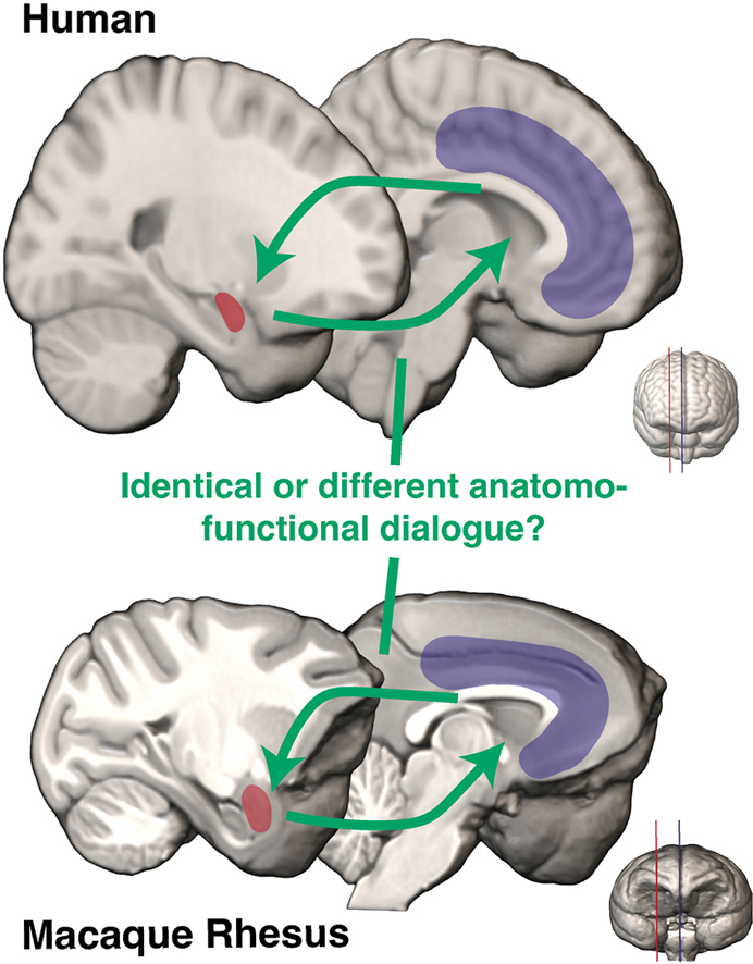
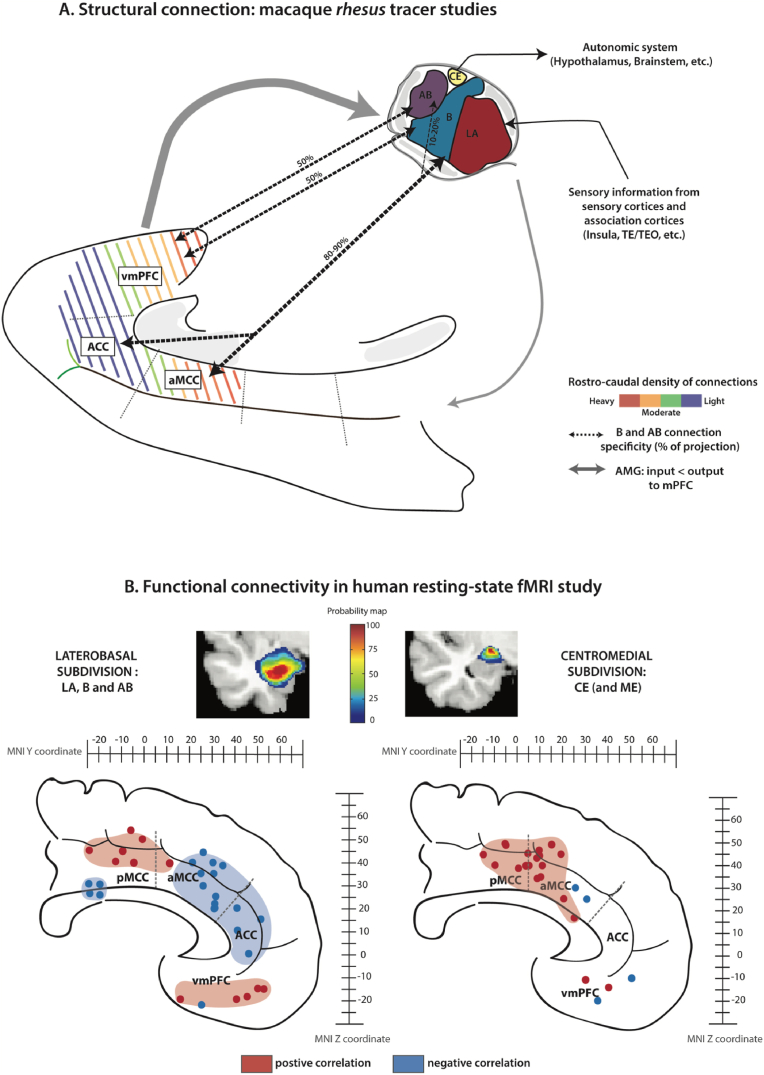
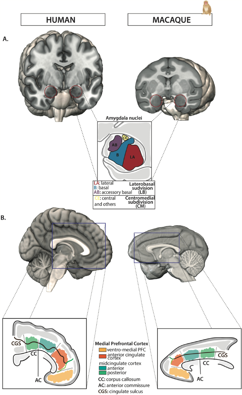
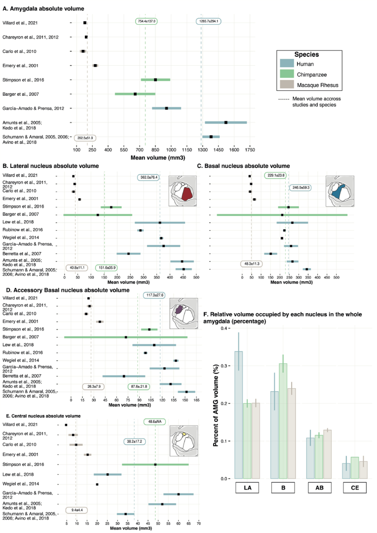
The network formed by the amygdala (AMG) and the medial Prefrontal Cortex (mPFC), at the interface between our internal and external environment, has been shown to support some important aspects of behavioral adaptation. Whether and how the anatomo-functional organization of this network evolved across primates remains unclear. Here, we compared AMG nuclei morphological characteristics and their functional connectivity with the mPFC in humans and macaques to identify potential homologies and differences between these species. Based on selected studies, we highlight two subsystems within the AMG-mPFC circuits, likely involved in distinct temporal dynamics of integration during behavioral adaptation. We also show that whereas the mPFC displays a large expansion but a preserved intrinsic anatomo-functional organization, the AMG displays a volume reduction and morphological changes related to specific nuclei. We discuss potential commonalities and differences in the dialogue between AMG nuclei and mPFC in humans and macaques based on available data.

摘要

杏仁核(AMG)和内侧前额叶皮质(mPFC)在我们的内部和外部环境之间的界面处形成的网络,已被证明支持行为适应的一些重要方面。该网络的解剖功能组织是否以及如何在灵长类动物中进化仍不清楚。在这里,我们比较了人类和猕猴中AMG核的形态特征及其与mPFC的功能连接,以确定这些物种之间潜在的同源性和差异。基于选定的研究,我们强调了AMG-mPFC回路中的两个子系统,它们可能参与行为适应过程中不同的时间整合动态。我们还表明,虽然mPFC显示出大规模扩张但保留了内在的解剖功能组织,而AMG则显示出体积减小和与特定核相关的形态变化。我们根据现有数据讨论了人类和猕猴中AMG核与mPFC之间对话中潜在的共性和差异。

https://cdn.ncbi.nlm.nih.gov/pmc/blobs/12fb/10432920/87d7c9aeda43/gr3.jpg
https://cdn.ncbi.nlm.nih.gov/pmc/blobs/12fb/10432920/6b2462c817ee/ga1.jpg
https://cdn.ncbi.nlm.nih.gov/pmc/blobs/12fb/10432920/430dc738111e/gr1.jpg
https://cdn.ncbi.nlm.nih.gov/pmc/blobs/12fb/10432920/e2c2f82dedb4/gr2.jpg
https://cdn.ncbi.nlm.nih.gov/pmc/blobs/12fb/10432920/87d7c9aeda43/gr3.jpg
https://cdn.ncbi.nlm.nih.gov/pmc/blobs/12fb/10432920/6b2462c817ee/ga1.jpg
https://cdn.ncbi.nlm.nih.gov/pmc/blobs/12fb/10432920/430dc738111e/gr1.jpg
https://cdn.ncbi.nlm.nih.gov/pmc/blobs/12fb/10432920/e2c2f82dedb4/gr2.jpg
https://cdn.ncbi.nlm.nih.gov/pmc/blobs/12fb/10432920/87d7c9aeda43/gr3.jpg

相似文献

1
Multiple routes of communication within the amygdala-mPFC network: A comparative approach in humans and macaques.杏仁核-内侧前额叶皮层网络内的多种通信途径:人类和猕猴的比较研究方法。
Curr Res Neurobiol. 2023 Jul 28;5:100103. doi: 10.1016/j.crneur.2023.100103. eCollection 2023.
2
Differential functional organization of amygdala-medial prefrontal cortex networks in macaque and human.杏仁核-前额叶皮质网络在猕猴和人类中的功能差异组织。
Commun Biol. 2024 Mar 5;7(1):269. doi: 10.1038/s42003-024-05918-y.
3
Two Days' Sleep Debt Causes Mood Decline During Resting State Via Diminished Amygdala-Prefrontal Connectivity.两天的睡眠债通过杏仁核-前额叶连接性减弱导致静息状态下情绪下降。
Sleep. 2017 Oct 1;40(10). doi: 10.1093/sleep/zsx133.
4
Endogenous cortisol is associated with functional connectivity between the amygdala and medial prefrontal cortex.内源性皮质醇与杏仁核和内侧前额叶皮质之间的功能连接有关。
Psychoneuroendocrinology. 2012 Jul;37(7):1039-47. doi: 10.1016/j.psyneuen.2011.12.001. Epub 2011 Dec 26.
5
Childhood Trauma and Functional Connectivity between Amygdala and Medial Prefrontal Cortex: A Dynamic Functional Connectivity and Large-Scale Network Perspective.童年创伤与杏仁核和内侧前额叶皮质之间的功能连接:动态功能连接和大规模网络视角
Front Syst Neurosci. 2017 May 11;11:29. doi: 10.3389/fnsys.2017.00029. eCollection 2017.
6
Frequency dependent emotion differentiation and directional coupling in amygdala, orbitofrontal and medial prefrontal cortex network with intracranial recordings.颅内记录显示杏仁核、眶额皮质和内侧前额叶皮质网络中的频率相关情绪分化和定向耦合。
Mol Psychiatry. 2023 Apr;28(4):1636-1646. doi: 10.1038/s41380-022-01883-2. Epub 2022 Dec 2.
7
Disconnection Between Amygdala and Medial Prefrontal Cortex in Psychotic Disorders.精神障碍中杏仁核与内侧前额叶皮质之间的联系中断
Schizophr Bull. 2016 Jul;42(4):1056-67. doi: 10.1093/schbul/sbw012. Epub 2016 Feb 23.
8
Heart rate variability is associated with amygdala functional connectivity with MPFC across younger and older adults.心率变异性与年轻人和老年人杏仁核与内侧前额叶皮质的功能连接有关。
Neuroimage. 2016 Oct 1;139:44-52. doi: 10.1016/j.neuroimage.2016.05.076. Epub 2016 May 31.
9
Organization of connections between the amygdala, medial prefrontal cortex, and lateral hypothalamus: a single and double retrograde tracing study in rats.杏仁核、内侧前额叶皮质和外侧下丘脑之间连接的组织架构:大鼠的单重和双重逆行追踪研究
Brain Struct Funct. 2016 Jul;221(6):2937-62. doi: 10.1007/s00429-015-1081-0. Epub 2015 Jul 14.
10
Optogenetic Examination of Prefrontal-Amygdala Synaptic Development.前额叶-杏仁核突触发育的光遗传学检测
J Neurosci. 2017 Mar 15;37(11):2976-2985. doi: 10.1523/JNEUROSCI.3097-16.2017. Epub 2017 Feb 13.

引用本文的文献

1
Hippocampal connectivity patterns echo macroscale cortical evolution in the primate brain.海马体连接模式在灵长类大脑中反映了宏观皮层的进化。
Nat Commun. 2024 Jul 16;15(1):5963. doi: 10.1038/s41467-024-49823-8.
2
Human resilience depends on distinctively human brain circuitry and development.人类的复原力取决于独特的人类大脑回路和发育。
Front Behav Neurosci. 2024 May 16;18:1370551. doi: 10.3389/fnbeh.2024.1370551. eCollection 2024.
3
Brainwide mesoscale functional networks revealed by focal infrared neural stimulation of the amygdala.

本文引用的文献

1
Amygdala circuit transitions supporting developmentally-appropriate social behavior.支持发展适当社会行为的杏仁核回路转变。
Neurobiol Learn Mem. 2023 May;201:107762. doi: 10.1016/j.nlm.2023.107762. Epub 2023 Apr 26.
2
Contributions of human amygdala nuclei to resting-state networks.人类杏仁核核团对静息态网络的贡献。
PLoS One. 2022 Dec 28;17(12):e0278962. doi: 10.1371/journal.pone.0278962. eCollection 2022.
3
Disorganized functional architecture of amygdala subregional networks in obsessive-compulsive disorder.强迫症患者杏仁核亚区网络功能结构紊乱。
杏仁核局部红外神经刺激揭示的全脑介观功能网络
bioRxiv. 2024 Jun 6:2024.02.14.580397. doi: 10.1101/2024.02.14.580397.
4
Differential functional organization of amygdala-medial prefrontal cortex networks in macaque and human.杏仁核-前额叶皮质网络在猕猴和人类中的功能差异组织。
Commun Biol. 2024 Mar 5;7(1):269. doi: 10.1038/s42003-024-05918-y.
Commun Biol. 2022 Nov 4;5(1):1184. doi: 10.1038/s42003-022-04115-z.
4
Relationship between nuclei-specific amygdala connectivity and mental health dimensions in humans.人类核内杏仁体连接与心理健康维度之间的关系。
Nat Hum Behav. 2022 Dec;6(12):1705-1722. doi: 10.1038/s41562-022-01434-3. Epub 2022 Sep 22.
5
The central extended amygdala guides survival-relevant tradeoffs: Implications for understanding common psychiatric disorders.中央延伸杏仁核指导与生存相关的权衡:对理解常见精神障碍的意义。
Neurosci Biobehav Rev. 2022 Nov;142:104879. doi: 10.1016/j.neubiorev.2022.104879. Epub 2022 Sep 15.
6
Mapping effective connectivity of human amygdala subdivisions with intracranial stimulation.颅内刺激下人类杏仁核亚区的有效连接图谱
Nat Commun. 2022 Aug 20;13(1):4909. doi: 10.1038/s41467-022-32644-y.
7
Altered resting state dynamic functional connectivity of amygdala subregions in patients with autism spectrum disorder: A multi-site fMRI study.孤独症谱系障碍患者杏仁核亚区静息态功能连接的改变:一项多中心 fMRI 研究。
J Affect Disord. 2022 Sep 1;312:69-77. doi: 10.1016/j.jad.2022.06.011. Epub 2022 Jun 13.
8
Widespread implementations of interactive social gaze neurons in the primate prefrontal-amygdala networks.普遍存在于灵长类动物前额叶-杏仁核网络中的交互社交凝视神经元。
Neuron. 2022 Jul 6;110(13):2183-2197.e7. doi: 10.1016/j.neuron.2022.04.013. Epub 2022 May 10.
9
Ultra-high field (10.5T) diffusion-weighted MRI of the macaque brain.恒磁场强度为 10.5T 的猕猴大脑弥散加权磁共振成像。
Neuroimage. 2022 Jul 15;255:119200. doi: 10.1016/j.neuroimage.2022.119200. Epub 2022 Apr 13.
10
Post mortem mapping of connectional anatomy for the validation of diffusion MRI.弥散磁共振成像连接解剖学的死后映射验证。
Neuroimage. 2022 Aug 1;256:119146. doi: 10.1016/j.neuroimage.2022.119146. Epub 2022 Mar 25.