1] Cancer Research Center, Sanford-Burnham Medical Research Institute, La Jolla, California 92037, USA [2] Center for Nanomedicine and Department of Chemistry and Biochemistry, University of California, Santa Barbara, California 93106, USA.
Cancer Research Center, Sanford-Burnham Medical Research Institute, La Jolla, California 92037, USA.
Nat Mater. 2014 Sep;13(9):904-11. doi: 10.1038/nmat3982. Epub 2014 Jun 8.
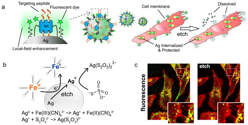
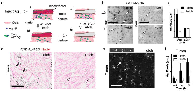
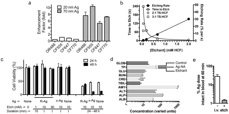
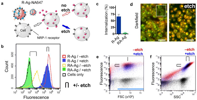
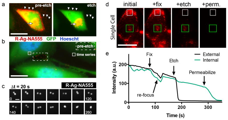
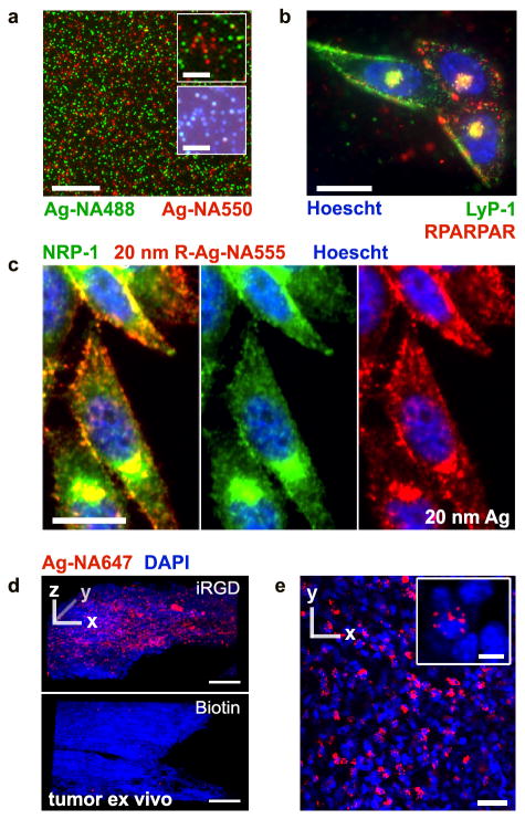
There is considerable interest in using nanoparticles as labels or to deliver drugs and other bioactive compounds to cells in vitro and in vivo. Fluorescent imaging, commonly used to study internalization and subcellular localization of nanoparticles, does not allow unequivocal distinction between cell surface-bound and internalized particles, as there is no methodology to turn particles 'off'. We have developed a simple technique to rapidly remove silver nanoparticles outside living cells, leaving only the internalized pool for imaging or quantification. The silver nanoparticle (AgNP) etching is based on the sensitivity of Ag to a hexacyanoferrate-thiosulphate redox-based destain solution. In demonstration of the technique we present a class of multicoloured plasmonic nanoprobes comprising dye-labelled AgNPs that are exceptionally bright and photostable, carry peptides as model targeting ligands, can be etched rapidly and with minimal toxicity in mice, and that show tumour uptake in vivo.
人们对使用纳米粒子作为标记物或向细胞内输送药物和其他生物活性化合物非常感兴趣,无论是在体外还是体内。荧光成像通常用于研究纳米粒子的内化和亚细胞定位,但由于没有将粒子“关闭”的方法,因此无法明确区分细胞表面结合的和内化的粒子。我们开发了一种简单的技术,可以快速去除活细胞外的银纳米粒子,只留下可用于成像或定量的内化池。银纳米粒子(AgNP)的蚀刻基于 Ag 对基于六氰合铁(II)-硫代硫酸盐的氧化还原去色溶液的敏感性。在技术演示中,我们提出了一类多色等离子体纳米探针,包括染料标记的 AgNPs,它们异常明亮且稳定,携带肽作为模型靶向配体,可以快速蚀刻,在小鼠体内具有最小的毒性,并且可以在体内显示肿瘤摄取。