Dumont Matthieu F, Yadavilli Sridevi, Sze Raymond W, Nazarian Javad, Fernandes Rohan
Sheikh Zayed Institute for Pediatric Surgical Innovation, Washington, DC, USA.
Center for Genetic Medicine Research, Children's National Medical Center, Washington, DC, USA.
Int J Nanomedicine. 2014 May 23;9:2581-95. doi: 10.2147/IJN.S63472. eCollection 2014.
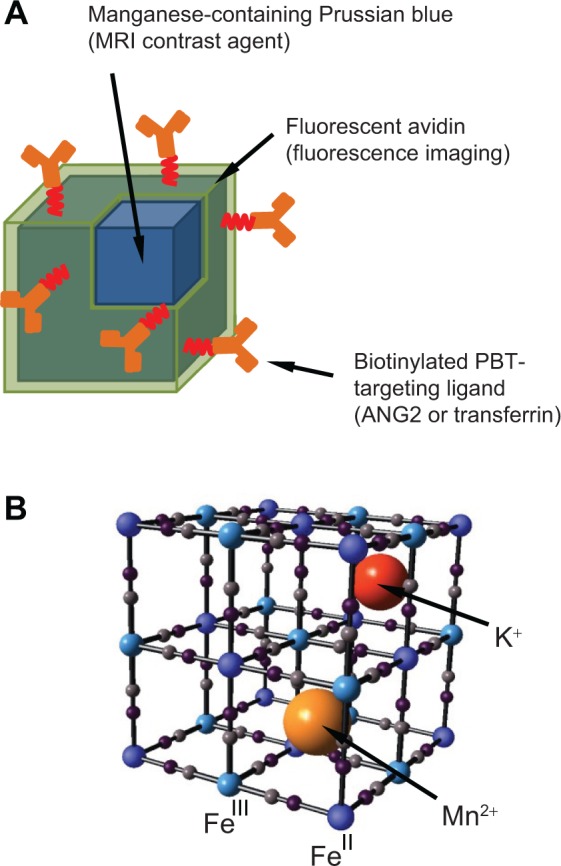
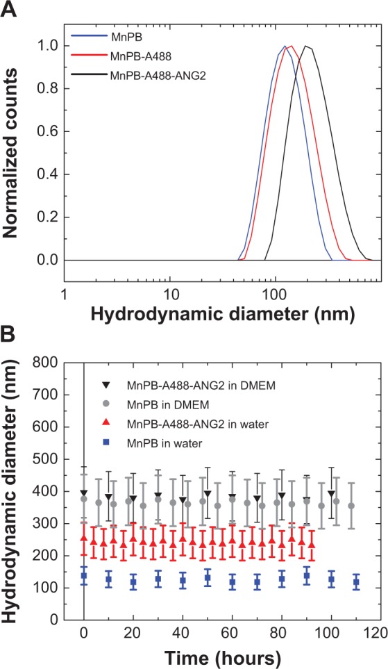
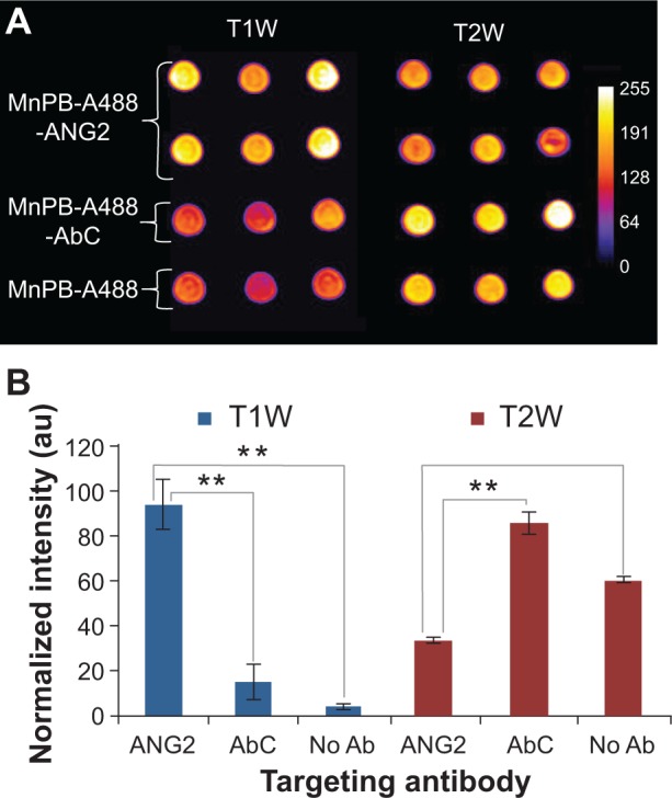
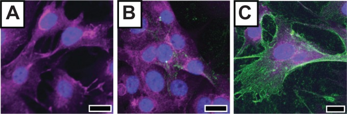
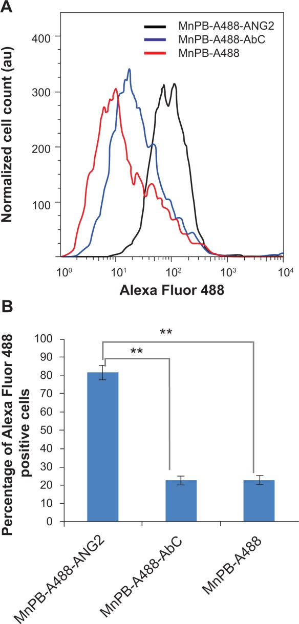
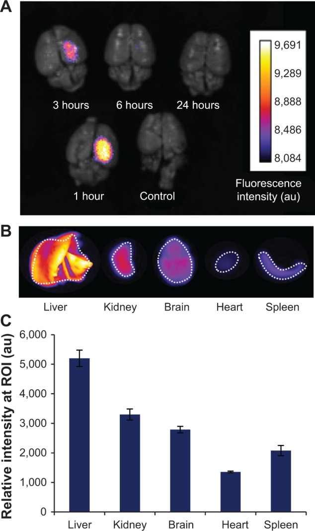
Pediatric brain tumors (PBTs) are a leading cause of death in children. For an improved prognosis in patients with PBTs, there is a critical need to develop molecularly-specific imaging agents to monitor disease progression and response to treatment. In this paper, we describe manganese-containing Prussian blue nanoparticles as agents for molecular magnetic resonance imaging (MRI) and fluorescence-based imaging of PBTs. Our core-shell nanoparticles consist of a core lattice structure that incorporates and retains paramagnetic Mn(2+) ions, and generates MRI contrast (both negative and positive). The biofunctionalized shell is comprised of fluorescent avidin, which serves the dual purpose of enabling fluorescence imaging and functioning as a platform for the attachment of biotinylated ligands that target PBTs. The surfaces of our nanoparticles are modified with biotinylated antibodies targeting neuron-glial antigen 2 or biotinylated transferrin. Both neuron-glial antigen 2 and the transferrin receptor are protein markers overexpressed in PBTs. We describe the synthesis, biofunctionalization, and characterization of these multimodal nanoparticles. Further, we demonstrate the MRI and fluorescence imaging capabilities of manganese-containing Prussian blue nanoparticles in vitro. Finally, we demonstrate the potential of these nanoparticles as PBT imaging agents by measuring their organ and brain biodistribution in an orthotopic mouse model of PBTs using ex vivo fluorescence imaging.
小儿脑肿瘤(PBTs)是儿童死亡的主要原因之一。为了改善PBTs患者的预后,迫切需要开发分子特异性成像剂来监测疾病进展和治疗反应。在本文中,我们描述了含锰普鲁士蓝纳米颗粒作为用于PBTs分子磁共振成像(MRI)和基于荧光成像的试剂。我们的核壳纳米颗粒由包含并保留顺磁性Mn(2+)离子的核心晶格结构组成,并产生MRI对比(包括阴性和阳性)。生物功能化外壳由荧光抗生物素蛋白组成,其具有双重作用,既能实现荧光成像,又能作为靶向PBTs的生物素化配体附着平台。我们纳米颗粒的表面用靶向神经胶质抗原2的生物素化抗体或生物素化转铁蛋白进行修饰。神经胶质抗原2和转铁蛋白受体都是在PBTs中过表达的蛋白质标志物。我们描述了这些多模态纳米颗粒的合成、生物功能化和表征。此外,我们在体外展示了含锰普鲁士蓝纳米颗粒的MRI和荧光成像能力。最后,我们通过使用离体荧光成像测量它们在PBTs原位小鼠模型中的器官和脑生物分布,证明了这些纳米颗粒作为PBT成像剂的潜力。