Barbaresi Paolo, Fabri Mara, Mensà Emanuela
Section of Neuroscience and Cell Biology, Department of Experimental and Clinical Medicine, Marche Polytechnic University Ancona, I-60020, Italy.
Brain Behav. 2014 May;4(3):317-36. doi: 10.1002/brb3.218. Epub 2014 Feb 12.
The aim of this study was to determine the presence and distribution of nitric oxide (NO)-producing neurons in the rat corpus callosum (cc).
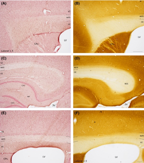
To investigate this aspect of cc organization we used nicotinamide adenine dinucleotide phosphate diaphorase (NADPH-d) histochemistry and neuronal NO synthase (nNOS) immunocytochemistry.
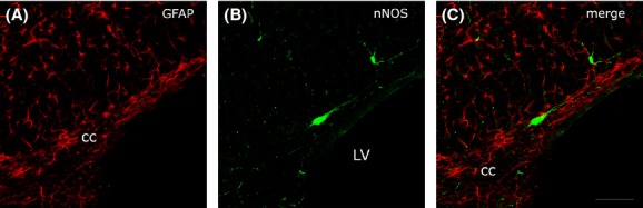
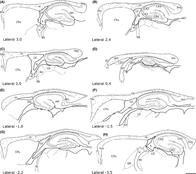
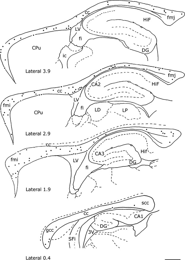
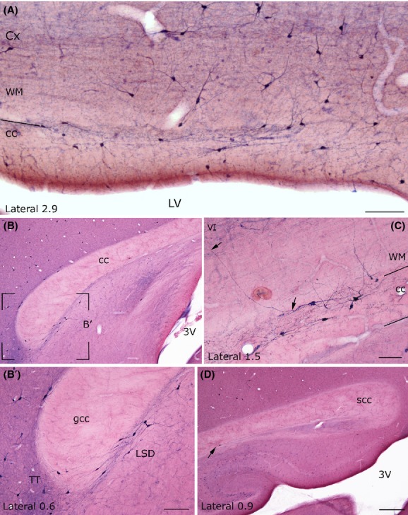
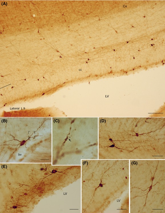
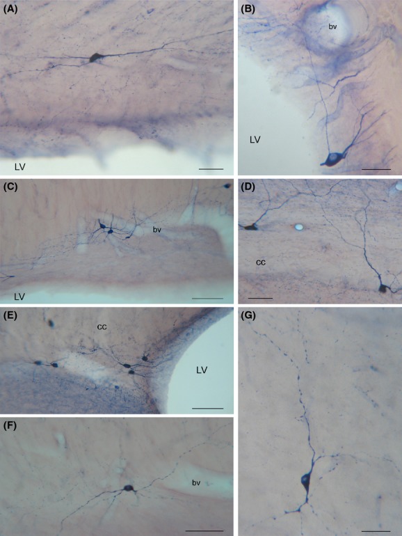
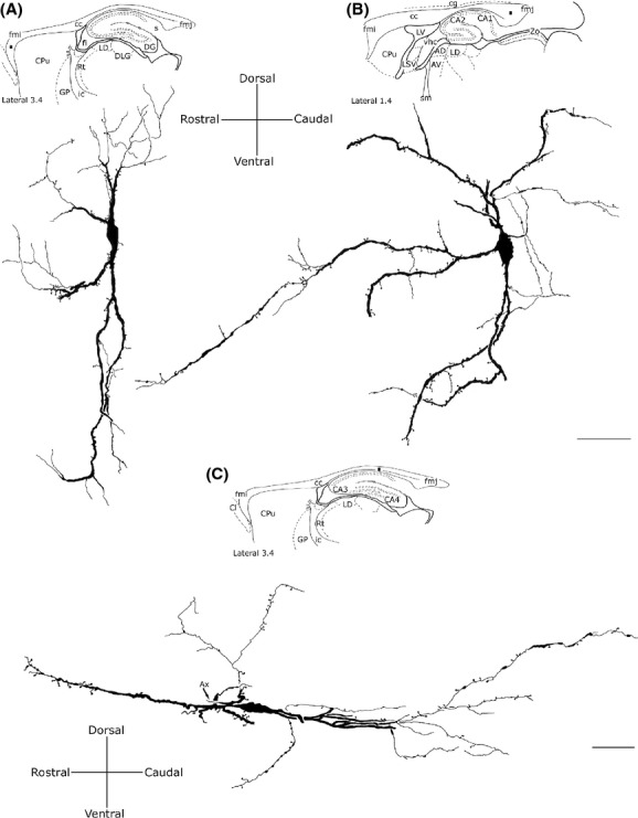
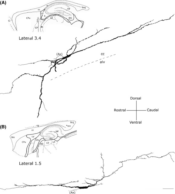
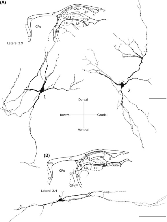
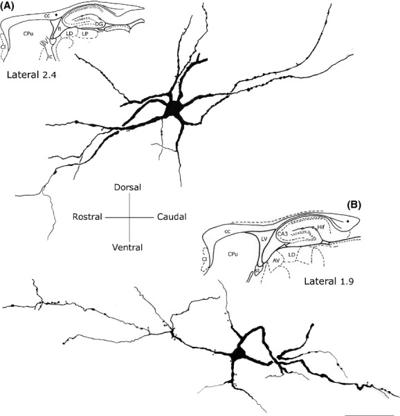
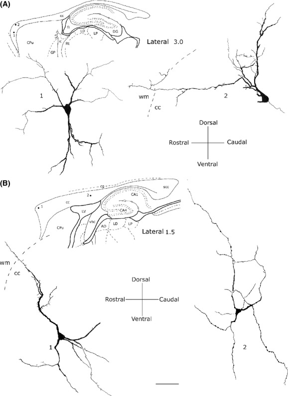
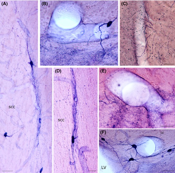
Intense NADPH-d-positive (NADPH-d+) neurons were found along the rostrocaudal extension of the cc (sagittal sections). They were more numerous in the lateral cc and gradually decreased in the more medial regions, where they were very few or absent. The Golgi-like appearance of NADPH-d+ intracallosal neurons allowed dividing them into five morphological types: (1) bipolar; (2) fusiform; (3) round; (4) polygonal; and (5) pyramidal. The number of NADPH-d+ neurons (both hemispheres) was counted in two brains using 50-μm thick sections. In the first brain, counts involved 145 sections and neurons were 2959; in the second, 2227 neurons were counted in 130 sections. The distribution and morphology of nNOS-immunopositive (nNOSIP) neurons was identical to that of NADPH-d+neurons. Some of these neurons were observed in the cc ependymal region, where they might be in contact with cerebrospinal fluid (CSF), monitoring its composition, pH, and osmolality changes, or playing a role in regulating the synthesis and release of several peptides. The somatic, dendritic, and axonal processes of many NADPH-d+/nNOSIP neurons were closely associated with intracallosal blood vessels.
Such close relationship raises the possibility that these neurons are a major source of NO during neural activity. As NO is a potent vasodilator, these findings strongly suggest that NO-positive neurons transduce neuronal signals into vascular responses in selected cc regions, thus giving rise to hemodynamic changes detectable by neuroimaging.
本研究的目的是确定大鼠胼胝体(cc)中产生一氧化氮(NO)的神经元的存在及其分布。
为了研究cc组织的这一方面,我们使用了烟酰胺腺嘌呤二核苷酸磷酸黄递酶(NADPH-d)组织化学和神经元型一氧化氮合酶(nNOS)免疫细胞化学方法。
在cc的前后延伸部分(矢状切片)发现了大量NADPH-d阳性(NADPH-d+)神经元。它们在cc外侧较多,在内侧区域逐渐减少,内侧区域数量很少或不存在。NADPH-d+胼胝体内神经元呈现出类似高尔基体的外观,可分为五种形态类型:(1)双极型;(2)梭形;(3)圆形;(4)多边形;(5)锥体形。使用50μm厚的切片在两个大脑中对NADPH-d+神经元(双侧半球)的数量进行了计数。在第一个大脑中,计数涉及145个切片,神经元数量为2959个;在第二个大脑中,在130个切片中计数到2227个神经元。nNOS免疫阳性(nNOSIP)神经元的分布和形态与NADPH-d+神经元相同。在cc室管膜区域观察到其中一些神经元,它们可能与脑脊液(CSF)接触,监测其成分、pH值和渗透压变化,或在调节几种肽的合成和释放中发挥作用。许多NADPH-d+/nNOSIP神经元的胞体、树突和轴突与胼胝体内血管密切相关。
这种密切关系增加了这些神经元在神经活动期间是NO主要来源的可能性。由于NO是一种强效血管舒张剂,这些发现强烈表明NO阳性神经元将神经元信号转化为特定cc区域的血管反应,从而引起神经影像学可检测到的血流动力学变化。