• 文献检索
  • 文档翻译
  • 深度研究
  • 学术资讯
  • Suppr Zotero 插件Zotero 插件
  • 邀请有礼
  • 套餐&价格
  • 历史记录
应用&插件
Suppr Zotero 插件Zotero 插件浏览器插件Mac 客户端Windows 客户端微信小程序
定价
高级版会员购买积分包购买API积分包
服务
文献检索文档翻译深度研究API 文档MCP 服务
关于我们
关于 Suppr公司介绍联系我们用户协议隐私条款
关注我们

Suppr 超能文献

核心技术专利:CN118964589B侵权必究
粤ICP备2023148730 号-1Suppr @ 2026

文献检索

告别复杂PubMed语法,用中文像聊天一样搜索,搜遍4000万医学文献。AI智能推荐,让科研检索更轻松。

立即免费搜索

文件翻译

保留排版,准确专业,支持PDF/Word/PPT等文件格式,支持 12+语言互译。

免费翻译文档

深度研究

AI帮你快速写综述,25分钟生成高质量综述,智能提取关键信息,辅助科研写作。

立即免费体验

光学显微镜下的倾斜视图重建。黑腹果蝇胚胎细胞核的三维重建。

Tilted view reconstruction in optical microscopy. Three-dimensional reconstruction of Drosophila melanogaster embryo nuclei.

作者信息

Shaw P J, Agard D A, Hiraoka Y, Sedat J W

机构信息

Department of Cell Biology, John Innes Institute, Norwich, United Kingdom.

出版信息

Biophys J. 1989 Jan;55(1):101-10. doi: 10.1016/S0006-3495(89)82783-3.

DOI:10.1016/S0006-3495(89)82783-3
PMID:2495031
原文链接:https://pmc.ncbi.nlm.nih.gov/articles/PMC1330446/
Abstract

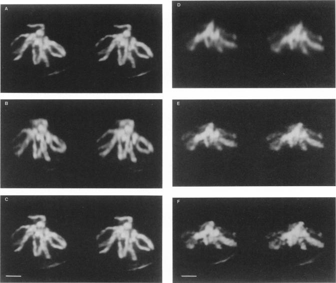
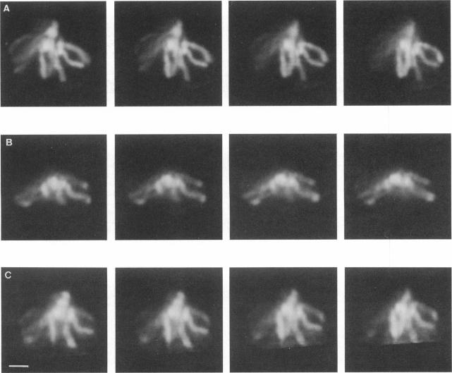
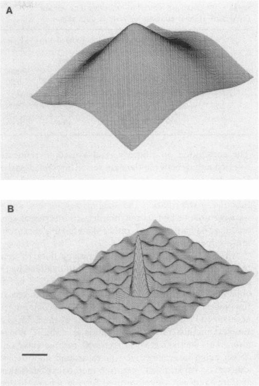
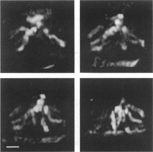
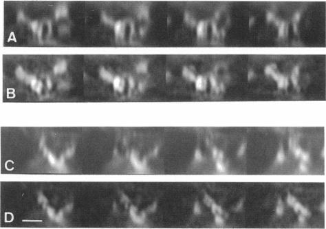
The resolution along the optical axis (z) is much less than the in-plane resolution in any current optical microscope, conventional or otherwise. We have used mutually tilted, through-focal section views of the same object to provide a solution to this problem. A tilting specimen stage was constructed for an optical microscope, which with the use of a coverslip-free water immersion lens, allowed the collection of data sets from intact Drosophila melanogaster embryos at viewing directions up to 90 degrees apart. We have devised an image processing scheme to determine the relative tilt, translation, and sampling parameters of the different data sets. This involves the use of a modified phase cross-correlation function, which produces a very sharp maximum. Finally the data sets are merged using figure-of-merit and local area scaling techniques borrowed from x-ray protein crystallography. We demonstrate the application of this technique to data sets from a metaphase plate in an embryo of Drosophila melanogaster. As expected, the merged reconstruction combined the highest resolution available in the individual data sets. As estimated from the Fourier transform, the final resolution is 0.25 microns in x and y and 0.4 microns in z. In the final reconstruction all ten chromosome arms can be easily delineated; this was not possible in the individual data sets. Within many of the arms the two individual chromatids can be seen. In some cases the chromatids are wrapped around each other helically, in others they lie alongside each other in a parallel arrangement.

摘要

在任何当前的光学显微镜中,无论是传统的还是其他类型的,沿光轴(z)方向的分辨率都远低于平面内分辨率。我们利用对同一物体相互倾斜的焦平面截面视图来解决这个问题。为光学显微镜构建了一个倾斜载物台,使用无盖玻片的水浸透镜,能够从完整的黑腹果蝇胚胎中以相差达90度的观察方向收集数据集。我们设计了一种图像处理方案来确定不同数据集的相对倾斜、平移和采样参数。这涉及使用一种改进的相位互相关函数,该函数会产生一个非常尖锐的最大值。最后,利用从X射线蛋白质晶体学借鉴的品质因数和局部区域缩放技术将数据集合并。我们展示了该技术在黑腹果蝇胚胎中期板数据集上的应用。正如预期的那样,合并后的重建结合了各个数据集中可用的最高分辨率。根据傅里叶变换估计,最终分辨率在x和y方向为0.25微米,在z方向为0.4微米。在最终重建中,可以轻松勾勒出所有十条染色体臂;这在单个数据集中是不可能的。在许多染色体臂内,可以看到两条单独的染色单体。在某些情况下,染色单体相互螺旋缠绕,在其他情况下,它们相互平行排列。

https://cdn.ncbi.nlm.nih.gov/pmc/blobs/48d1/1330446/7a73512ea496/biophysj00145-0114-a.jpg
https://cdn.ncbi.nlm.nih.gov/pmc/blobs/48d1/1330446/e3cd59d75bc0/biophysj00145-0108-a.jpg
https://cdn.ncbi.nlm.nih.gov/pmc/blobs/48d1/1330446/467cad701a63/biophysj00145-0110-a.jpg
https://cdn.ncbi.nlm.nih.gov/pmc/blobs/48d1/1330446/42990ac5eed1/biophysj00145-0111-a.jpg
https://cdn.ncbi.nlm.nih.gov/pmc/blobs/48d1/1330446/2f9ae20ca8bf/biophysj00145-0112-a.jpg
https://cdn.ncbi.nlm.nih.gov/pmc/blobs/48d1/1330446/79cb1592e2a2/biophysj00145-0113-a.jpg
https://cdn.ncbi.nlm.nih.gov/pmc/blobs/48d1/1330446/6b7e67e093a5/biophysj00145-0113-b.jpg
https://cdn.ncbi.nlm.nih.gov/pmc/blobs/48d1/1330446/7a73512ea496/biophysj00145-0114-a.jpg

相似文献

1
Tilted view reconstruction in optical microscopy. Three-dimensional reconstruction of Drosophila melanogaster embryo nuclei.光学显微镜下的倾斜视图重建。黑腹果蝇胚胎细胞核的三维重建。
Biophys J. 1989 Jan;55(1):101-10. doi: 10.1016/S0006-3495(89)82783-3.
2
Three-dimensional optical microscopy using tilted views.使用倾斜视图的三维光学显微镜术。
J Microsc. 1990 May;158(Pt 2):165-72. doi: 10.1111/j.1365-2818.1990.tb02989.x.
3
A tilting device for three-dimensional microscopy: application to in situ imaging of interphase cell nuclei.一种用于三维显微镜检查的倾斜装置:在间期细胞核原位成像中的应用。
J Microsc. 1992 Oct;168(Pt 1):47-57. doi: 10.1111/j.1365-2818.1992.tb03249.x.
4
Superresolution measurements in vivo: imaging Drosophila embryo by photoactivated localization microscopy.体内超分辨率测量:通过光激活定位显微镜对果蝇胚胎进行成像
Methods Cell Biol. 2015;125:119-42. doi: 10.1016/bs.mcb.2014.10.015. Epub 2015 Jan 8.
5
Direct cell lineage analysis in Drosophila melanogaster by time-lapse, three-dimensional optical microscopy of living embryos.通过对活体胚胎进行延时三维光学显微镜观察,对黑腹果蝇进行直接细胞谱系分析。
J Cell Biol. 1989 Aug;109(2):505-16. doi: 10.1083/jcb.109.2.505.
6
High-resolution microscopic methods for the analysis of cellular movements in Drosophila embryos.用于分析果蝇胚胎细胞运动的高分辨率显微方法。
Methods Cell Biol. 1994;44:507-32. doi: 10.1016/s0091-679x(08)60929-2.
7
Interpretation of the three-dimensional structure of living nuclei by specimen tilt.
J Cell Sci. 1975 Oct;19(1):1-10. doi: 10.1242/jcs.19.1.1.
8
Three-color immunofluorescence imaging of Drosophila embryos by laser scanning confocal microscopy.通过激光扫描共聚焦显微镜对果蝇胚胎进行三色免疫荧光成像。
Biotechniques. 1993 Jan;14(1):42-8.
9
Light-microscope specimen holder with 3-axis rotation and small-angle control.带有三轴旋转和小角度控制的光学显微镜标本载物台。
J Neurosci Methods. 2014 Jan 15;221:15-21. doi: 10.1016/j.jneumeth.2013.08.026. Epub 2013 Sep 8.
10
Dual-axis tomography: an approach with alignment methods that preserve resolution.双轴断层扫描:一种采用保持分辨率的对齐方法的技术。
J Struct Biol. 1997 Dec;120(3):343-52. doi: 10.1006/jsbi.1997.3919.

引用本文的文献

1
Fast and noise-tolerant determination of the center of rotation in tomography.断层扫描中旋转中心的快速且抗噪声确定
J Synchrotron Radiat. 2022 Mar 1;29(Pt 2):488-495. doi: 10.1107/S1600577521012777. Epub 2022 Jan 19.
2
Multiview deconvolution approximation multiphoton microscopy of tissues and zebrafish larvae.多视图去卷积近似多光子显微镜技术在组织和斑马鱼幼体中的应用。
Sci Rep. 2021 May 12;11(1):10160. doi: 10.1038/s41598-021-89566-w.
3
Multiview microscopy of single cells through microstructure-based indirect optical manipulation.

本文引用的文献

1
Optical sectioning microscopy: cellular architecture in three dimensions.光学切片显微镜术:三维细胞结构
Annu Rev Biophys Bioeng. 1984;13:191-219. doi: 10.1146/annurev.bb.13.060184.001203.
2
Three-dimensional architecture of a polytene nucleus.多线染色体核的三维结构。
Nature. 1983 Apr 21;302(5910):676-81. doi: 10.1038/302676a0.
3
Localization of antigenic determinants in whole Drosophila embryos.果蝇全胚胎中抗原决定簇的定位
通过基于微观结构的间接光学操控实现单细胞的多视角显微镜观察。
Biomed Opt Express. 2020 Jan 16;11(2):945-962. doi: 10.1364/BOE.379233. eCollection 2020 Feb 1.
4
High-resolution ultramicroscopy of the developing and adult nervous system in optically cleared Drosophila melanogaster.在透明化的黑腹果蝇的发育中和成年神经系统中的高分辨率超微结构研究。
Nat Commun. 2018 Nov 9;9(1):4731. doi: 10.1038/s41467-018-07192-z.
5
Multidirectional Image Sensing for Microscopy Based on a Rotatable Robot.基于可旋转机器人的显微镜多向图像传感
Sensors (Basel). 2015 Dec 15;15(12):31566-80. doi: 10.3390/s151229872.
6
Controlled 3D rotation of biological cells using optical multiple-force clamps.利用光学多力钳对生物细胞进行可控的三维旋转。
Biomed Opt Express. 2014 Jun 18;5(7):2341-8. doi: 10.1364/BOE.5.002341. eCollection 2014 Jul 1.
7
Going "open" with mesoscopy: a new dimension on multi-view imaging.将腹腔镜检查技术应用于“开放手术”:多视角成像的新维度。
Protoplasma. 2014 Mar;251(2):363-72. doi: 10.1007/s00709-013-0599-3. Epub 2014 Jan 18.
8
Correlative microscopy methods that maximize specimen fidelity and data completeness, and improve molecular localization capabilities.最大化标本保真度和数据完整性,并提高分子定位能力的相关显微镜方法。
J Struct Biol. 2013 Oct;184(1):12-20. doi: 10.1016/j.jsb.2013.03.006. Epub 2013 Mar 24.
9
Optical Tomography in weakly scattering media in the presence of highly scattering inclusions.存在高散射内含物时弱散射介质中的光学层析成像。
Biomed Opt Express. 2011 Jan 31;2(3):440-51. doi: 10.1364/BOE.2.000440.
10
Selective plane illumination microscopy techniques in developmental biology.发育生物学中的选择性平面照明显微技术。
Development. 2009 Jun;136(12):1963-75. doi: 10.1242/dev.022426.
Dev Biol. 1983 Sep;99(1):261-4. doi: 10.1016/0012-1606(83)90275-0.
4
A simple chamber for observing microscopic specimens in both top and side views.一个用于从顶部和侧面观察微观标本的简易腔室。
J Microsc. 1985 Jan;137(Pt 1):29-34. doi: 10.1111/j.1365-2818.1985.tb02558.x.
5
The use of a charge-coupled device for quantitative optical microscopy of biological structures.电荷耦合器件在生物结构定量光学显微镜检查中的应用。
Science. 1987 Oct 2;238(4823):36-41. doi: 10.1126/science.3116667.
6
An evaluation of confocal versus conventional imaging of biological structures by fluorescence light microscopy.通过荧光显微镜对生物结构进行共聚焦成像与传统成像的评估。
J Cell Biol. 1987 Jul;105(1):41-8. doi: 10.1083/jcb.105.1.41.
7
Motif detection in quantum noise-limited electron micrographs by cross-correlation.通过互相关检测量子噪声限制电子显微照片中的基序
Ultramicroscopy. 1977 Apr;2(2-3):219-27. doi: 10.1016/s0304-3991(76)91385-1.
8
Interpretation of the three-dimensional structure of living nuclei by specimen tilt.
J Cell Sci. 1975 Oct;19(1):1-10. doi: 10.1242/jcs.19.1.1.