Hirata Akira, Yamamoto Soichiro, Okinami Satoshi
Department of Ophthalmology, Faculty of Medicine, Saga University, 5-1-1, Nabeshima, Saga 849-8501, Japan.
J Funct Biomater. 2013 Jan 18;4(1):6-13. doi: 10.3390/jfb4010006.
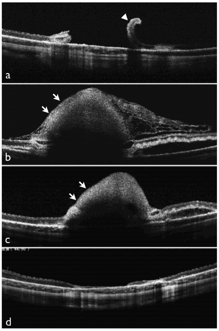
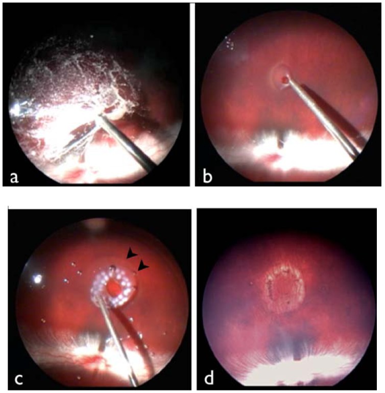
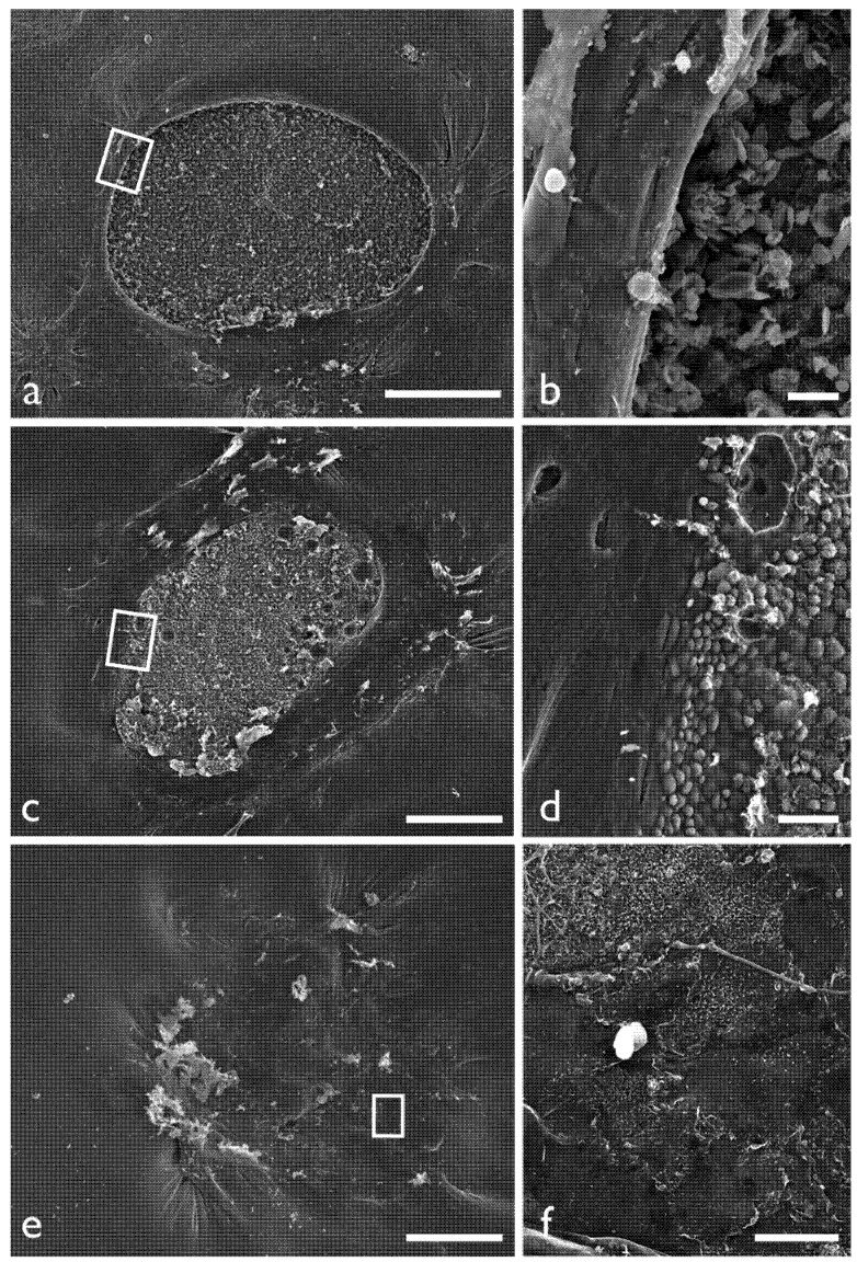
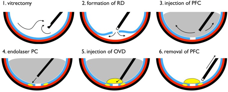
To investigate the temporary tamponade effects of an ophthalmic viscosurgical device (OVD) for experimental retinal tears, we performed vitrectomy in four rabbit eyes and created a posterior vitreous detachment and artificial retinal tear to produce retinal detachment. The retina was flattened with liquid perfluorocarbon (PFC), the area peripheral to the tear was photocoagulated, an OVD was applied to the retinal tear surface below the PFC and the PFC was removed by aspiration. In the control group, PFC was removed without application of OVD. At one, three and seven days postoperatively, funduscopy and optical coherence tomography (OCT) were performed to examine the sealing process of the retinal tear. In OVD-treated eyes, the OVD remained on the retinal surface, and the retinal tear was patched for ≥ 3 days postoperatively. By seven days postoperatively, the OVD on the retinal surface had disappeared, and the retina was reattached. In control eyes, the edge of the retinal tear was rolled, and retinal detachment persisted. In OVD-treated eyes, the border of the retinal tear was indistinct, and the defect area was significantly decreased. These results show that application of an OVD effectively seals retinal tears and eliminates retinal detachments.
为研究眼科粘弹剂(OVD)对实验性视网膜裂孔的临时填塞效果,我们对4只兔眼进行了玻璃体切除术,造成玻璃体后脱离和人工视网膜裂孔以导致视网膜脱离。用全氟碳化物(PFC)液体使视网膜变平,对裂孔周围区域进行光凝,将OVD应用于PFC下方的视网膜裂孔表面,然后通过抽吸去除PFC。在对照组中,去除PFC但不应用OVD。术后1天、3天和7天,进行眼底镜检查和光学相干断层扫描(OCT)以检查视网膜裂孔的封闭过程。在接受OVD治疗的眼中,OVD保留在视网膜表面,视网膜裂孔在术后≥3天得到修补。术后7天时,视网膜表面的OVD消失,视网膜重新附着。在对照眼中,视网膜裂孔边缘卷曲,视网膜脱离持续存在。在接受OVD治疗的眼中,视网膜裂孔边界不清,缺损区域明显减小。这些结果表明,应用OVD可有效封闭视网膜裂孔并消除视网膜脱离。