Walk Elyse L, McLaughlin Sarah, Coad James, Weed Scott A
Department of Neurobiology and Anatomy, West Virginia University, Morgantown, West Virginia, United States of America; Program in Cancer Cell Biology, West Virginia University, Morgantown, West Virginia, United States of America; Mary Babb Randolph Cancer Center, West Virginia University, Morgantown, West Virginia, United States of America.
Animal Models and Imaging Facility, West Virginia University, Morgantown, West Virginia, United States of America; Mary Babb Randolph Cancer Center, West Virginia University, Morgantown, West Virginia, United States of America.
PLoS One. 2014 Jun 23;9(6):e100185. doi: 10.1371/journal.pone.0100185. eCollection 2014.
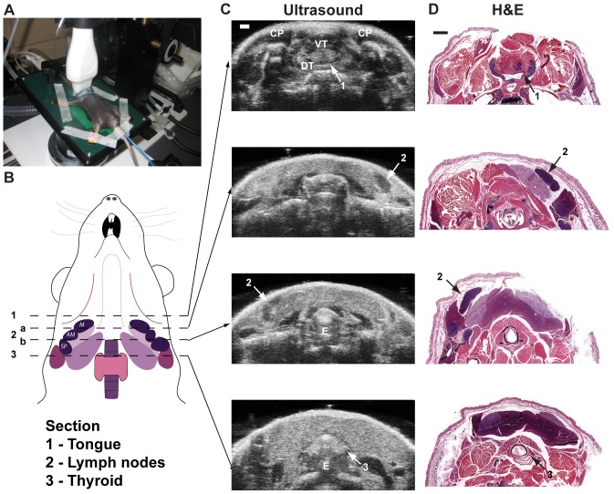
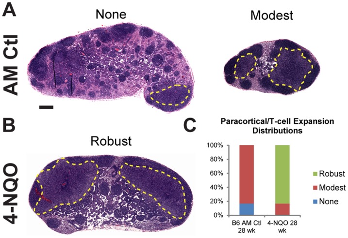
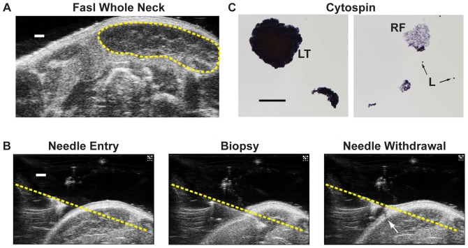
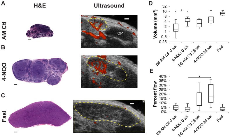
Cervical lymph node evaluation by clinical ultrasound is a non-invasive procedure used in diagnosing nodal status, and when combined with fine-needle aspiration cytology (FNAC), provides an effective method to assess nodal pathologies. Development of high-frequency ultrasound (HF US) allows real-time monitoring of lymph node alterations in animal models. While HF US is frequently used in animal models of tumor biology, use of HF US for studying cervical lymph nodes alterations associated with murine models of head and neck cancer, or any other model of lymphadenopathy, is lacking. Here we utilize HF US to monitor cervical lymph nodes changes in mice following exposure to the oral cancer-inducing carcinogen 4-nitroquinoline-1-oxide (4-NQO) and in mice with systemic autoimmunity. 4-NQO induces tumors within the mouse oral cavity as early as 19 wks that recapitulate HNSCC. Monitoring of cervical (mandibular) lymph nodes by gray scale and power Doppler sonography revealed changes in lymph node size eight weeks after 4-NQO treatment, prior to tumor formation. 4-NQO causes changes in cervical node blood flow resulting from oral tumor progression. Histological evaluation indicated that the early 4-NQO induced changes in lymph node volume were due to specific hyperproliferation of T-cell enriched zones in the paracortex. We also show that HF US can be used to perform image-guided fine needle aspirate (FNA) biopsies on mice with enlarged mandibular lymph nodes due to genetic mutation of Fas ligand (Fasl). Collectively these studies indicate that HF US is an effective technique for the non-invasive study of cervical lymph node alterations in live mouse models of oral cancer and other mouse models containing cervical lymphadenopathy.
通过临床超声评估颈部淋巴结是一种用于诊断淋巴结状态的非侵入性程序,与细针穿刺细胞学检查(FNAC)相结合时,可提供一种评估淋巴结病变的有效方法。高频超声(HF US)的发展使得在动物模型中能够实时监测淋巴结的变化。虽然HF US在肿瘤生物学动物模型中经常使用,但缺乏将HF US用于研究与头颈癌小鼠模型或任何其他淋巴结病模型相关的颈部淋巴结变化的研究。在这里,我们利用HF US监测小鼠在暴露于口腔癌诱导致癌物4-硝基喹啉-1-氧化物(4-NQO)后以及患有系统性自身免疫的小鼠的颈部淋巴结变化。4-NQO最早在19周时在小鼠口腔内诱发肿瘤,重现头颈部鳞状细胞癌(HNSCC)。通过灰阶和能量多普勒超声对颈部(下颌)淋巴结进行监测,发现在4-NQO治疗8周后,即在肿瘤形成之前,淋巴结大小发生了变化。4-NQO导致口腔肿瘤进展引起颈部淋巴结血流变化。组织学评估表明,4-NQO早期诱导的淋巴结体积变化是由于副皮质中富含T细胞区域的特异性过度增殖所致。我们还表明,HF US可用于对因Fas配体(Fasl)基因突变而导致下颌淋巴结肿大的小鼠进行图像引导下的细针抽吸(FNA)活检。总的来说,这些研究表明,HF US是一种用于在口腔癌活体小鼠模型和其他含有颈部淋巴结病的小鼠模型中对颈部淋巴结变化进行非侵入性研究的有效技术。