• 文献检索
  • 文档翻译
  • 深度研究
  • 学术资讯
  • Suppr Zotero 插件Zotero 插件
  • 邀请有礼
  • 套餐&价格
  • 历史记录
应用&插件
Suppr Zotero 插件Zotero 插件浏览器插件Mac 客户端Windows 客户端微信小程序
定价
高级版会员购买积分包购买API积分包
服务
文献检索文档翻译深度研究API 文档MCP 服务
关于我们
关于 Suppr公司介绍联系我们用户协议隐私条款
关注我们

Suppr 超能文献

核心技术专利:CN118964589B侵权必究
粤ICP备2023148730 号-1Suppr @ 2026

文献检索

告别复杂PubMed语法,用中文像聊天一样搜索,搜遍4000万医学文献。AI智能推荐,让科研检索更轻松。

立即免费搜索

文件翻译

保留排版,准确专业,支持PDF/Word/PPT等文件格式,支持 12+语言互译。

免费翻译文档

深度研究

AI帮你快速写综述,25分钟生成高质量综述,智能提取关键信息,辅助科研写作。

立即免费体验

CT及PET/CT成像观察孤立性薄壁空洞型肺癌的表现及病理特征

Manifestations and pathological features of solitary thin-walled cavity lung cancer observed by CT and PET/CT imaging.

作者信息

Qi Yuangang, Zhang Qing, Huang Yong, Wang Daoqing

机构信息

Department of Radiology, Affiliated Hospital of Shandong Academy of Medical Sciences, Jinan, Shandong 250032, P.R. China.

Department of Radiology, Shandong Cancer Hospital, Jinan, Shandong 250114, P.R. China.

出版信息

Oncol Lett. 2014 Jul;8(1):285-290. doi: 10.3892/ol.2014.2065. Epub 2014 Apr 15.

DOI:10.3892/ol.2014.2065
PMID:24959262
原文链接:https://pmc.ncbi.nlm.nih.gov/articles/PMC4063653/
Abstract

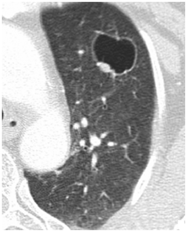
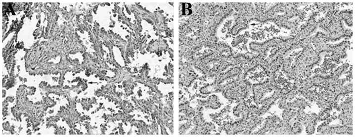
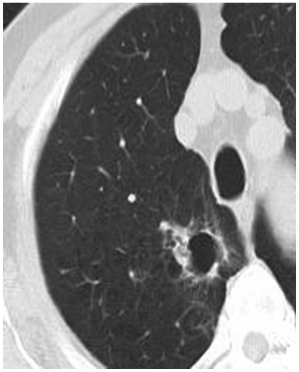
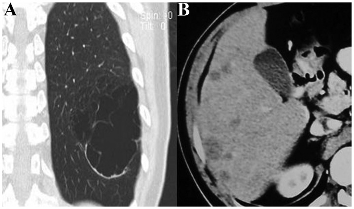
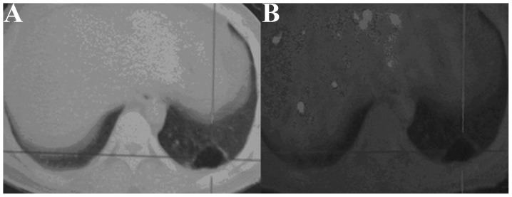
The aim of the present study was to analyze and improve the understanding of computed tomography (CT) and positron emission tomography (PET)/CT imaging and the pathological features of solitary thin-walled cavity lung cancer. A total of 16 patients with pathologically confirmed solitary thin-walled cavity lung cancer were included in the present study. All of the patients received CT scans. Among these, two patients underwent an additional PET/CT examination. The CT and PET/CT images were analyzed and a cross-check analysis of the pathological results was conducted. In total, 16 cases of lesions demonstrated thin-walled cavities on the CT images. Among these cases, three presented with an uneven thickening of the cavity walls, 10 cases exhibited wall nodules and three cases presented with compartments in the cavity. The standard uptake value (SUV) of the cavity wall increased in two patients who underwent PET/CT examinations. The 16 cases of lesions were pathologically confirmed as adenocarcinomas. Light microscopy revealed that the tumor cells, which were observed in 12 cases of lesions, had diffused along the inner cavity wall and the tumor cells of four cases had invaded the bronchial wall. Images of the chest that demonstrated a single thin-walled cavity accompanied by uneven thickening of the cavity wall or wall nodules, in addition to an increase in the SUV and compartments in the cavity, indicated potential lung cancer. Valves formed as a result of bronchial wall damage may have led to the cavity.

摘要

本研究的目的是分析并增进对计算机断层扫描(CT)和正电子发射断层扫描(PET)/CT成像以及孤立性薄壁空洞型肺癌病理特征的理解。本研究共纳入16例经病理证实的孤立性薄壁空洞型肺癌患者。所有患者均接受了CT扫描。其中,2例患者还接受了PET/CT检查。对CT和PET/CT图像进行了分析,并对病理结果进行了交叉核对分析。总共16例病变在CT图像上显示为薄壁空洞。在这些病例中,3例表现为空洞壁增厚不均匀,10例有壁结节,3例有空洞内分隔。接受PET/CT检查的2例患者空洞壁的标准化摄取值(SUV)升高。16例病变经病理证实为腺癌。光学显微镜检查显示,12例病变中观察到的肿瘤细胞沿内腔壁扩散,4例病变的肿瘤细胞侵犯了支气管壁。胸部图像显示单个薄壁空洞,伴有空洞壁增厚不均匀或壁结节,此外SUV升高和有空洞内分隔,提示可能为肺癌。支气管壁损伤形成的活瓣可能导致了空洞的形成。

https://cdn.ncbi.nlm.nih.gov/pmc/blobs/2543/4063653/d012292f9133/OL-08-01-0285-g06.jpg
https://cdn.ncbi.nlm.nih.gov/pmc/blobs/2543/4063653/a8aeffecb262/OL-08-01-0285-g00.jpg
https://cdn.ncbi.nlm.nih.gov/pmc/blobs/2543/4063653/44d0b663173d/OL-08-01-0285-g01.jpg
https://cdn.ncbi.nlm.nih.gov/pmc/blobs/2543/4063653/9914ac8c9149/OL-08-01-0285-g02.jpg
https://cdn.ncbi.nlm.nih.gov/pmc/blobs/2543/4063653/1a403ada6e13/OL-08-01-0285-g03.jpg
https://cdn.ncbi.nlm.nih.gov/pmc/blobs/2543/4063653/cc77f4dae790/OL-08-01-0285-g04.jpg
https://cdn.ncbi.nlm.nih.gov/pmc/blobs/2543/4063653/78b4298715ea/OL-08-01-0285-g05.jpg
https://cdn.ncbi.nlm.nih.gov/pmc/blobs/2543/4063653/d012292f9133/OL-08-01-0285-g06.jpg

相似文献

1
Manifestations and pathological features of solitary thin-walled cavity lung cancer observed by CT and PET/CT imaging.CT及PET/CT成像观察孤立性薄壁空洞型肺癌的表现及病理特征
Oncol Lett. 2014 Jul;8(1):285-290. doi: 10.3892/ol.2014.2065. Epub 2014 Apr 15.
2
Comparative study of solitary thin-walled cavity lung cancer with computed tomography and pathological findings.孤立性薄壁空洞型肺癌的 CT 与病理对照研究。
Lung Cancer. 2012 Oct;78(1):45-50. doi: 10.1016/j.lungcan.2012.06.004. Epub 2012 Jul 10.
3
Computed tomography for the diagnosis of solitary thin-walled cavity lung cancer.计算机断层扫描在孤立性薄壁空洞型肺癌诊断中的应用
Clin Respir J. 2015 Oct;9(4):392-8. doi: 10.1111/crj.12172. Epub 2014 Jul 23.
4
CT imaging features of thin-walled cavitary squamous cell lung cancer.薄壁空洞型肺鳞状细胞癌的CT影像特征
Adv Respir Med. 2019;87(2):114-117. doi: 10.5603/ARM.2019.0018.
5
Cavity Wall Thickness in Solitary Cavitary Lung Adenocarcinomas Is a Prognostic Indicator.孤立性空洞型肺腺癌的空洞壁厚度是一个预后指标。
Ann Thorac Surg. 2016 Dec;102(6):1863-1871. doi: 10.1016/j.athoracsur.2016.03.121. Epub 2016 Sep 20.
6
[Combined use of thin-section CT and F-FDG PET/CT for characterization of solitary pulmonary nodules].[薄层CT与F-FDG PET/CT联合应用于孤立性肺结节的特征分析]
Nan Fang Yi Ke Da Xue Xue Bao. 2017 Mar 20;37(3):283-289. doi: 10.3969/j.issn.1673-4254.2017.03.01.
7
Respiratory-gated imaging in metabolic evaluation of small solitary pulmonary nodules: 18F-FDG PET/CT and correlation with histology.呼吸门控成像在小的孤立性肺结节代谢评估中的应用:18F-FDG PET/CT及其与组织学的相关性
Nucl Med Commun. 2015 Jul;36(7):722-7. doi: 10.1097/MNM.0000000000000311.
8
Dual time point fluorodeoxyglucose positron emission tomography/computed tomography in differentiation between malignant and benign lesions in cancer patients. Does it always work?双时间点氟脱氧葡萄糖正电子发射断层扫描/计算机断层扫描在癌症患者恶性与良性病变鉴别中的应用。它总能奏效吗?
Indian J Nucl Med. 2015 Oct-Dec;30(4):314-9. doi: 10.4103/0972-3919.159693.
9
More advantages in detecting bone and soft tissue metastases from prostate cancer using F-PSMA PET/CT.使用F-PSMA PET/CT检测前列腺癌骨和软组织转移方面有更多优势。
Hell J Nucl Med. 2019 Jan-Apr;22(1):6-9. doi: 10.1967/s002449910952. Epub 2019 Mar 7.
10
Accuracy of FDG PET/CT in the evaluation of solitary pulmonary lesions - own experience.FDG PET/CT在孤立性肺结节评估中的准确性——自身经验
Pneumonol Alergol Pol. 2014;82(3):198-205. doi: 10.5603/PiAP.2014.0027.

引用本文的文献

1
A Comprehensive Review of Advances in Molecular Mechanisms and Targeted Therapies for the Specific Type of Cystic Lung Cancer.特定类型囊性肺癌的分子机制与靶向治疗进展综述
Onco Targets Ther. 2025 Feb 11;18:211-224. doi: 10.2147/OTT.S495018. eCollection 2025.
2
[Clinical Multi-features Analysis of Cystic Lung Adenocarcinoma 
and Construction of Invasive Risk Prediction Model].[肺囊性腺癌的临床多特征分析及侵袭性风险预测模型的构建]
Zhongguo Fei Ai Za Zhi. 2024 Apr 20;27(4):266-275. doi: 10.3779/j.issn.1009-3419.2024.102.14.
3
Lung cancer with air lucency: a systematic review and clinical management guide.

本文引用的文献

1
Comparative study of solitary thin-walled cavity lung cancer with computed tomography and pathological findings.孤立性薄壁空洞型肺癌的 CT 与病理对照研究。
Lung Cancer. 2012 Oct;78(1):45-50. doi: 10.1016/j.lungcan.2012.06.004. Epub 2012 Jul 10.
2
[Synchronous multiple lung cancer including a lesion with a thin-walled cavity; report of a case].[同步性多发性肺癌,包括一个具有薄壁空洞的病灶;病例报告]
Kyobu Geka. 2010 Feb;63(2):164-7.
3
[An autopsied case of mucinous bronchioloalveolar carcinoma associated with multiple thin-walled cavities].
伴有空气透亮影的肺癌:一项系统综述及临床管理指南
J Thorac Dis. 2023 Feb 28;15(2):731-746. doi: 10.21037/jtd-22-1199. Epub 2023 Feb 23.
4
Pitfalls in the surgical treatment of undiagnosed lung lesions and cystic pulmonary hydatidosis.未确诊肺部病变和肺包虫囊肿病的外科治疗陷阱。
J Cardiothorac Surg. 2022 Oct 27;17(1):275. doi: 10.1186/s13019-022-02026-y.
5
Lung adenocarcinoma initially mimicking localized emphysema.最初表现为局限性肺气肿的肺腺癌
Transl Cancer Res. 2020 Mar;9(3):2074-2076. doi: 10.21037/tcr.2020.01.29.
6
Comparative study of imaging and pathological evaluation of pneumonic mucinous adenocarcinoma.肺炎性黏液腺癌的影像学与病理评估的对比研究
Oncol Lett. 2021 Feb;21(2):125. doi: 10.3892/ol.2020.12386. Epub 2020 Dec 17.
7
Congenital pulmonary airway malformation mimicking lung cancer: A case report.酷似肺癌的先天性肺气道畸形:一例报告。
Medicine (Baltimore). 2019 Jun;98(24):e16057. doi: 10.1097/MD.0000000000016057.
8
The mechanism of formation of thin-walled cystic lung cancer.薄壁囊性肺癌的形成机制。
Medicine (Baltimore). 2019 Apr;98(14):e15031. doi: 10.1097/MD.0000000000015031.
9
Solitary thin-walled cystic lung cancer with extensive extrapulmonary metastasis: A case report and review of the literature.孤立性薄壁囊性肺癌伴广泛肺外转移:一例报告并文献复习
Medicine (Baltimore). 2018 Oct;97(43):e12950. doi: 10.1097/MD.0000000000012950.
10
Thin-wall cystic lung cancer: A study of 45 cases.薄壁囊性肺癌:45例病例研究
Oncol Lett. 2018 Jul;16(1):755-760. doi: 10.3892/ol.2018.8707. Epub 2018 May 14.
[一例伴有多个薄壁空洞的黏液性细支气管肺泡癌尸检病例]
Nihon Kokyuki Gakkai Zasshi. 2009 Jun;47(6):512-7.
4
[A case of lung adenocarcinoma with gradual enlargement of thin-walled cavity causing pneumothorax].1例肺腺癌伴薄壁空洞逐渐增大致气胸
Nihon Kokyuki Gakkai Zasshi. 2008 Jul;46(7):552-7.
5
Computer-aided diagnosis in lung nodule assessment.肺结节评估中的计算机辅助诊断
J Thorac Imaging. 2008 May;23(2):97-104. doi: 10.1097/RTI.0b013e318173dd1f.
6
Percutaneous needle washing for the diagnosis of pulmonary thin-walled cavitary lesions filled with air.经皮针吸冲洗用于诊断肺部充满气体的薄壁空洞性病变。
Intern Med. 2007;46(14):1089-94. doi: 10.2169/internalmedicine.46.0020. Epub 2007 Jul 17.
7
PET/CT imaging of lung cancer.肺癌的正电子发射断层显像/X线计算机体层成像(PET/CT)检查
J Thorac Imaging. 2006 May;21(2):123-36. doi: 10.1097/00005382-200605000-00004.
8
CT screening for lung cancer: five-year prospective experience.CT筛查肺癌:五年前瞻性经验。
Radiology. 2005 Apr;235(1):259-65. doi: 10.1148/radiol.2351041662. Epub 2005 Feb 4.
9
Mediastinal lymph node involvement in potentially resectable lung cancer: comparison of CT, positron emission tomography, and endoscopic ultrasonography with and without fine-needle aspiration.潜在可切除肺癌的纵隔淋巴结受累情况:CT、正电子发射断层扫描以及有无细针穿刺的内镜超声检查的比较
Chest. 2003 Feb;123(2):442-51. doi: 10.1378/chest.123.2.442.
10
Lung nodule enhancement at CT: multicenter study.CT 上肺结节的强化:多中心研究。
Radiology. 2000 Jan;214(1):73-80. doi: 10.1148/radiology.214.1.r00ja1473.