Institute of Cell Signalling, School of Life Sciences, Medical School, and.
School of Pharmacy, Centre for Biomolecular Sciences, University of Nottingham, Nottingham, UK.
FASEB J. 2014 Oct;28(10):4211-22. doi: 10.1096/fj.13-247270. Epub 2014 Jun 26.
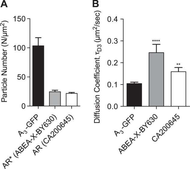
In our previous work, using a fluorescent adenosine-A3 receptor (A3AR) agonist and fluorescence correlation spectroscopy (FCS), we demonstrated high-affinity labeling of the active receptor (R*) conformation. In the current study, we used a fluorescent A3AR antagonist (CA200645) to study the binding characteristics of antagonist-occupied inactive receptor (R) conformations in membrane microdomains of individual cells. FCS analysis of CA200645-occupied A3ARs revealed 2 species, τD2 and τD3, that diffused at 2.29 ± 0.35 and 0.09 ± 0.03 μm(2)/s, respectively. FCS analysis of a green fluorescent protein (GFP)-tagged A3AR exhibited a single diffusing species (0.105 μm(2)/s). The binding of CA200645 to τD3 was antagonized by nanomolar concentrations of the A3 antagonist MRS 1220, but not by the agonist NECA (up to 300 nM), consistent with labeling of R. CA200645 normally dissociated slowly from the A3AR, but inclusion of xanthine amine congener (XAC) or VUF 5455 during washout markedly accelerated the reduction in the number of particles exhibiting τD3 characteristics. It is notable that this effect was accompanied by a significant increase in the number of particles with τD2 diffusion. These data show that FCS analysis of ligand-occupied receptors provides a unique means of monitoring ligand A3AR residence times that are significantly reduced as a consequence of allosteric interaction across the dimer interface
在我们之前的工作中,使用荧光腺苷-A3 受体 (A3AR) 激动剂和荧光相关光谱 (FCS),我们证明了高亲和力标记活性受体 (R*)构象。在当前的研究中,我们使用荧光 A3AR 拮抗剂 (CA200645) 来研究配体占据的失活受体 (R)构象在单个细胞的膜微域中的结合特性。CA200645 占据的 A3AR 的 FCS 分析显示出 2 种扩散,τD2 和 τD3,分别以 2.29 ± 0.35 和 0.09 ± 0.03 μm(2)/s 的速度扩散。绿色荧光蛋白 (GFP) 标记的 A3AR 的 FCS 分析显示出单一的扩散物种 (0.105 μm(2)/s)。CA200645 与 τD3 的结合被 A3 拮抗剂 MRS 1220 的纳摩尔浓度拮抗,但不受激动剂 NECA 的影响(高达 300 nM),与 R 的标记一致。CA200645 通常从 A3AR 缓慢解离,但在冲洗过程中包含黄嘌呤胺同系物 (XAC) 或 VUF 5455 时,显著加速了显示 τD3 特征的粒子数量的减少。值得注意的是,这种效应伴随着 τD2 扩散的粒子数量的显著增加。这些数据表明,配体占据的受体的 FCS 分析提供了一种独特的监测配体 A3AR 停留时间的方法,由于二聚体界面的变构相互作用,停留时间显著缩短。