de Jong Pim A, Hellings Willem E, Takx Richard A P, Išgum Ivana, van Herwaarden Joost A, Mali Willem P Th M
Department of Radiology, University Medical Center Utrecht, Utrecht, The Netherlands.
Department of Vascular Surgery, University Medical Center Utrecht, Utrecht, The Netherlands.
PLoS One. 2014 Jul 8;9(7):e102036. doi: 10.1371/journal.pone.0102036. eCollection 2014.
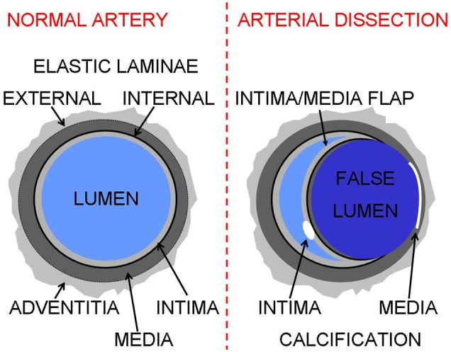
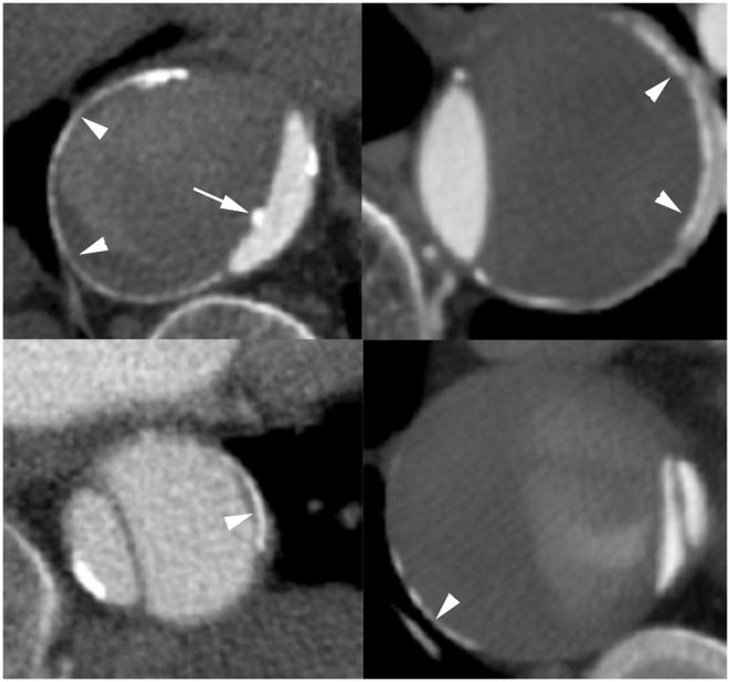
To investigate the frequency of aortic calcifications at the outer edge of the false lumen and the frequency of fully circular aortic calcifications in a consecutive series of patients with aortic dissection who underwent contrast-enhanced CT.
The study population compromised of 69 consecutive subjects aged 60 years and older with a contrast-enhanced CT scan demonstrating an aortic dissection. All CT scans were evaluated for the frequency of aortic calcifications at the outer edge of the false lumen and the frequency of fully circular aortic calcifications by two experienced observers. Between observer reliability was evaluated by using Cohen's Kappa. Differences between groups were tested using unpaired T test and Chi-square test.
Presumed media calcifications were observed in 22 (32%) patients of 60 years and older and were found more frequently in chronic aortic dissection (N = 12/23, 52%) than in acute aortic dissection (N = 10/46, 22%).
As the intima has been torn away by the aortic dissection it is highly likely that CT scans can visualize the calcifications in the tunica media of the aorta.
在一系列接受增强CT检查的主动脉夹层患者中,研究假腔外缘主动脉钙化的发生率以及完全环形主动脉钙化的发生率。
研究人群包括69例连续的60岁及以上接受增强CT扫描显示主动脉夹层的受试者。两名经验丰富的观察者对所有CT扫描进行评估,以确定假腔外缘主动脉钙化的发生率和完全环形主动脉钙化的发生率。观察者间的可靠性采用Cohen's Kappa进行评估。组间差异采用非配对t检验和卡方检验。
在60岁及以上的22例(32%)患者中观察到推测的中膜钙化,在慢性主动脉夹层(N = 12/23,52%)中比在急性主动脉夹层(N = 10/46,22%)中更常见。
由于主动脉夹层已将内膜撕裂,CT扫描极有可能显示主动脉中膜的钙化。