Department of Ophthalmology, Saga University Faculty of Medicine, 5-1-1, Nabeshima, Saga, 849-8501, Japan,
Ophthalmol Ther. 2013 Jun;2(1):11-8. doi: 10.1007/s40123-012-0008-6. Epub 2013 Jan 4.
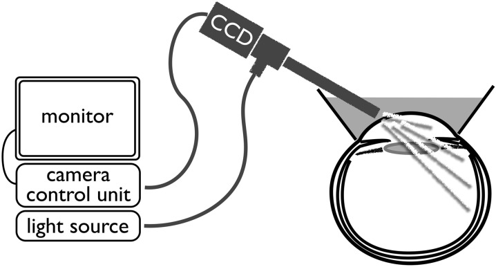
This study evaluated the usefulness of rigid endoscopy placed on the corneal surface to observe the peripheral retina.
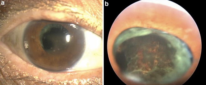
The authors studied 15 eyes in 15 patients (12 men, 3 women; mean age 55.9 years; range 22-74 years) that underwent vitreous surgery at the Department of Ophthalmology at Saga University Hospital. With patients in a supine position, after topical anesthesia, an eye cup was placed between the eyelids and filled with hydroxyethyl cellulose solution and physiologic saline. With a rigid endoscope placed near the corneal surface, the target areas were then observed and recorded. The usefulness of rigid endoscopy to observe the peripheral retina was evaluated based on differences due to lens status and pupil size.
In seven aphakic eyes, irrespective of pupil size, the peripheral retina could be observed up to the entire ora serrata (all quadrants). In eight eyes implanted with an intraocular lens, the observable area changed with pupil size and anterior capsulorrhexis size.
This technique using rigid endoscopy was simple to manipulate and useful for observing and recording the peripheral retina. In particular, in aphakic eyes, irrespective of pupil size, the retina could be observed to the ora serrata.
本研究评估了将硬性内镜置于角膜表面观察周边视网膜的有用性。
作者研究了在佐贺大学医院眼科接受玻璃体手术的 15 名患者(12 名男性,3 名女性;平均年龄 55.9 岁;范围 22-74 岁)的 15 只眼。患者取仰卧位,表面麻醉后,将眼杯置于眼睑之间,并用羟乙基纤维素溶液和生理盐水填充。将硬性内镜置于角膜表面附近,然后观察并记录目标区域。根据晶状体状态和瞳孔大小的差异评估硬性内镜观察周边视网膜的有用性。
在 7 只无晶状体眼中,无论瞳孔大小如何,周边视网膜均可观察到整个锯齿缘(所有象限)。在 8 只植入人工晶状体的眼中,可观察区域随瞳孔大小和前囊膜切开术大小而变化。
使用硬性内镜的这种技术操作简单,可用于观察和记录周边视网膜。特别是在无晶状体眼中,无论瞳孔大小如何,均可观察到锯齿缘。