• 文献检索
  • 文档翻译
  • 深度研究
  • 学术资讯
  • Suppr Zotero 插件Zotero 插件
  • 邀请有礼
  • 套餐&价格
  • 历史记录
应用&插件
Suppr Zotero 插件Zotero 插件浏览器插件Mac 客户端Windows 客户端微信小程序
定价
高级版会员购买积分包购买API积分包
服务
文献检索文档翻译深度研究API 文档MCP 服务
关于我们
关于 Suppr公司介绍联系我们用户协议隐私条款
关注我们

Suppr 超能文献

核心技术专利:CN118964589B侵权必究
粤ICP备2023148730 号-1Suppr @ 2026

文献检索

告别复杂PubMed语法,用中文像聊天一样搜索,搜遍4000万医学文献。AI智能推荐,让科研检索更轻松。

立即免费搜索

文件翻译

保留排版,准确专业,支持PDF/Word/PPT等文件格式,支持 12+语言互译。

免费翻译文档

深度研究

AI帮你快速写综述,25分钟生成高质量综述,智能提取关键信息,辅助科研写作。

立即免费体验

一种作为骨科植入材料的纳米羟基磷灰石/聚醚醚酮生物复合材料的制备、表征及体外成骨细胞功能

Preparation, characterization, and in vitro osteoblast functions of a nano-hydroxyapatite/polyetheretherketone biocomposite as orthopedic implant material.

作者信息

Ma Rui, Tang Songchao, Tan Honglue, Lin Wentao, Wang Yugang, Wei Jie, Zhao Liming, Tang Tingting

机构信息

Shanghai Key Laboratory of Orthopedic Implants, Department of Orthopedic Surgery, Shanghai Ninth People's Hospital, Shanghai Jiao Tong University School of Medicine, People's Republic of China.

Key Laboratory for Ultrafine Materials of Ministry of Education and The State Key Laboratory of Bioreactor Engineering, East China University of Science and Technology, Shanghai, People's Republic of China.

出版信息

Int J Nanomedicine. 2014 Aug 18;9:3949-61. doi: 10.2147/IJN.S67358. eCollection 2014.

DOI:10.2147/IJN.S67358
PMID:25170265
原文链接:https://pmc.ncbi.nlm.nih.gov/articles/PMC4145828/
Abstract

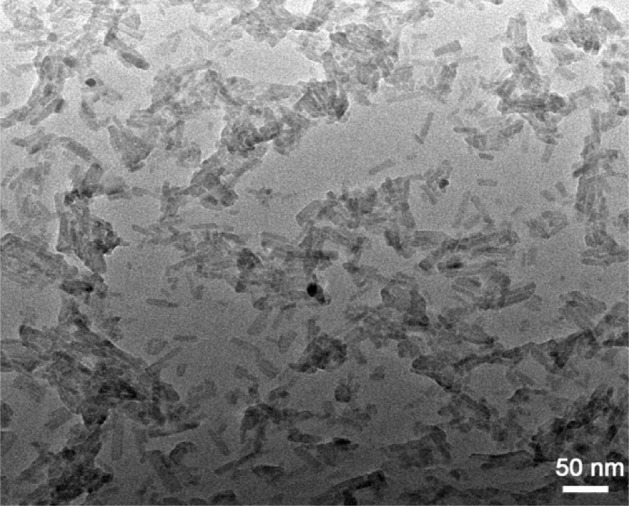
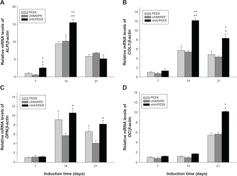
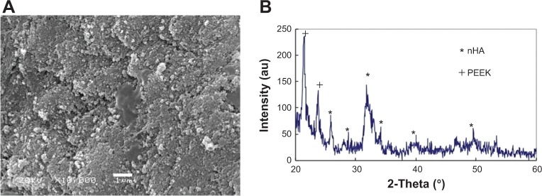
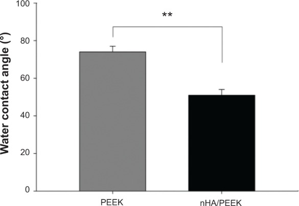
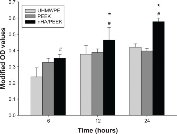
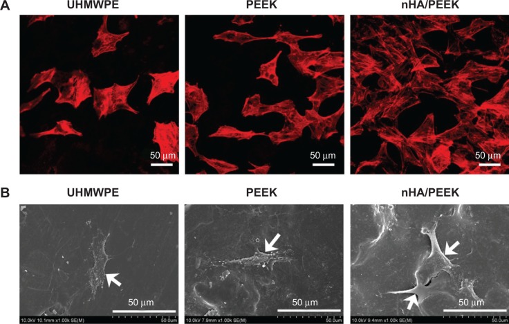
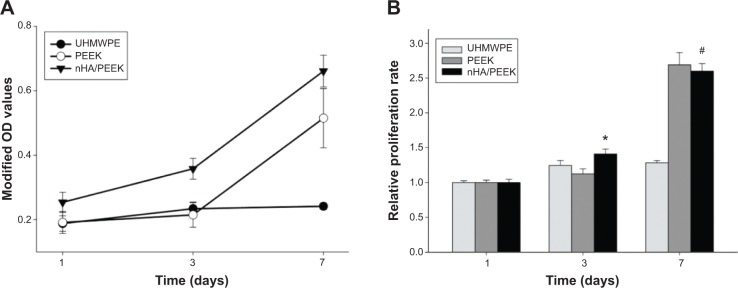
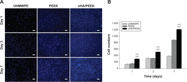
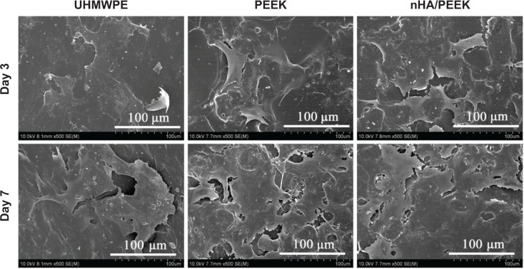
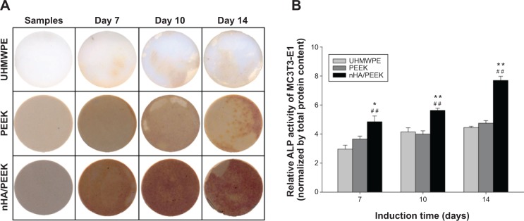
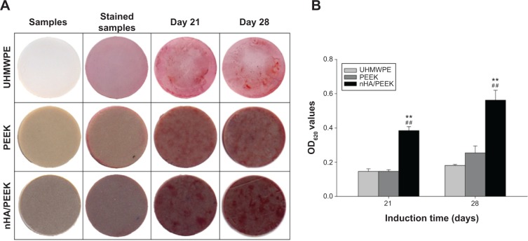
A bioactive composite was prepared by incorporating 40 wt% nano-hydroxyapatite (nHA) into polyetheretherketone (PEEK) through a process of compounding, injection, and molding. The mechanical and surface properties of the nHA/PEEK composite were characterized, and the in vitro osteoblast functions in the composite were investigated. The mechanical properties (elastic modulus and compressive strength) of the nHA/PEEK composite increased significantly, while the tensile strength decreased slightly as compared with PEEK. Further, the addition of nHA into PEEK increased the surface roughness and hydrophilicity of the nHA/PEEK composite. In cell tests, compared with PEEK and ultra-high-molecular-weight polyethylene, it was found that the nHA/PEEK composite could promote the functions of MC3T3-E1 cells, including cell attachment, spreading, proliferation, alkaline phosphatase activity, calcium nodule formation, and expression of osteogenic differentiation-related genes. Incorporation of nHA into PEEK greatly improved the bioperformance of PEEK. The nHA/PEEK composite might be a promising orthopedic implant material.

摘要

通过复合、注塑和成型工艺,将40重量%的纳米羟基磷灰石(nHA)掺入聚醚醚酮(PEEK)中制备了一种生物活性复合材料。对nHA/PEEK复合材料的力学性能和表面性能进行了表征,并研究了该复合材料在体外的成骨细胞功能。与PEEK相比,nHA/PEEK复合材料的力学性能(弹性模量和抗压强度)显著提高,而拉伸强度略有下降。此外,向PEEK中添加nHA增加了nHA/PEEK复合材料的表面粗糙度和亲水性。在细胞测试中,发现与PEEK和超高分子量聚乙烯相比,nHA/PEEK复合材料能够促进MC3T3-E1细胞的功能,包括细胞附着、铺展、增殖、碱性磷酸酶活性、钙结节形成以及成骨分化相关基因的表达。将nHA掺入PEEK中大大改善了PEEK的生物性能。nHA/PEEK复合材料可能是一种很有前途的骨科植入材料。

https://cdn.ncbi.nlm.nih.gov/pmc/blobs/0e48/4145828/78443dba051b/ijn-9-3949Fig11.jpg
https://cdn.ncbi.nlm.nih.gov/pmc/blobs/0e48/4145828/852b420763e3/ijn-9-3949Fig1.jpg
https://cdn.ncbi.nlm.nih.gov/pmc/blobs/0e48/4145828/08e40d66dea3/ijn-9-3949Fig2.jpg
https://cdn.ncbi.nlm.nih.gov/pmc/blobs/0e48/4145828/4458c84133bd/ijn-9-3949Fig3.jpg
https://cdn.ncbi.nlm.nih.gov/pmc/blobs/0e48/4145828/255ce5bde390/ijn-9-3949Fig4.jpg
https://cdn.ncbi.nlm.nih.gov/pmc/blobs/0e48/4145828/3eda3f99f828/ijn-9-3949Fig5.jpg
https://cdn.ncbi.nlm.nih.gov/pmc/blobs/0e48/4145828/7c1fe9dbdc18/ijn-9-3949Fig6.jpg
https://cdn.ncbi.nlm.nih.gov/pmc/blobs/0e48/4145828/1db51e5c5f3c/ijn-9-3949Fig7.jpg
https://cdn.ncbi.nlm.nih.gov/pmc/blobs/0e48/4145828/4240f7b6e5c6/ijn-9-3949Fig8.jpg
https://cdn.ncbi.nlm.nih.gov/pmc/blobs/0e48/4145828/92eec63e7fcb/ijn-9-3949Fig9.jpg
https://cdn.ncbi.nlm.nih.gov/pmc/blobs/0e48/4145828/1521e507b7b8/ijn-9-3949Fig10.jpg
https://cdn.ncbi.nlm.nih.gov/pmc/blobs/0e48/4145828/78443dba051b/ijn-9-3949Fig11.jpg

相似文献

1
Preparation, characterization, and in vitro osteoblast functions of a nano-hydroxyapatite/polyetheretherketone biocomposite as orthopedic implant material.一种作为骨科植入材料的纳米羟基磷灰石/聚醚醚酮生物复合材料的制备、表征及体外成骨细胞功能
Int J Nanomedicine. 2014 Aug 18;9:3949-61. doi: 10.2147/IJN.S67358. eCollection 2014.
2
Effect of surface roughness on osteogenesis in vitro and osseointegration in vivo of carbon fiber-reinforced polyetheretherketone-nanohydroxyapatite composite.表面粗糙度对碳纤维增强聚醚醚酮-纳米羟基磷灰石复合材料体外成骨及体内骨整合的影响
Int J Nanomedicine. 2015 Feb 17;10:1425-47. doi: 10.2147/IJN.S75557. eCollection 2015.
3
Enhanced osteogenic activity of phosphorylated polyetheretherketone via surface-initiated grafting polymerization of vinylphosphonic acid.通过表面引发接枝聚合乙烯基膦酸增强聚醚醚酮的成骨活性。
Colloids Surf B Biointerfaces. 2019 Jan 1;173:591-598. doi: 10.1016/j.colsurfb.2018.10.031. Epub 2018 Oct 13.
4
Preparation, characterization, cellular response and in vivo osseointegration of polyetheretherketone/nano-hydroxyapatite/carbon fiber ternary biocomposite.聚醚醚酮/纳米羟基磷灰石/碳纤维三元生物复合材料的制备、表征、细胞反应及体内骨整合
Colloids Surf B Biointerfaces. 2015 Dec 1;136:64-73. doi: 10.1016/j.colsurfb.2015.09.001. Epub 2015 Sep 2.
5
Nano-TiO2/PEEK bioactive composite as a bone substitute material: in vitro and in vivo studies.纳米 TiO2/PEEK 生物活性复合材料作为骨替代材料的体外与体内研究。
Int J Nanomedicine. 2012;7:1215-25. doi: 10.2147/IJN.S28101. Epub 2012 Mar 5.
6
Evaluating the bioactivity of a hydroxyapatite-incorporated polyetheretherketone biocomposite.评估含羟基磷灰石的聚醚醚酮生物复合材料的生物活性。
J Orthop Surg Res. 2019 Jan 25;14(1):32. doi: 10.1186/s13018-019-1069-1.
7
Preparation, mechanical properties and in vitro cytocompatibility of multi-walled carbon nanotubes/poly(etheretherketone) nanocomposites.多壁碳纳米管/聚醚醚酮纳米复合材料的制备、力学性能及体外细胞相容性。
J Biomater Sci Polym Ed. 2018 Mar;29(4):428-447. doi: 10.1080/09205063.2017.1422852. Epub 2018 Jan 9.
8
Preparation, characterization, in vitro bioactivity, and cellular responses to a polyetheretherketone bioactive composite containing nanocalcium silicate for bone repair.聚醚醚酮生物活性复合材料的制备、表征、体外生物活性及对骨修复的细胞反应,该复合材料含有纳米硅酸钙。
ACS Appl Mater Interfaces. 2014 Aug 13;6(15):12214-25. doi: 10.1021/am504409q. Epub 2014 Jul 24.
9
Enhancement of osteogenesis on micro/nano-topographical carbon fiber-reinforced polyetheretherketone-nanohydroxyapatite biocomposite.微/纳米拓扑结构碳纤维增强聚醚醚酮-纳米羟基磷灰石生物复合材料上骨生成的增强
Mater Sci Eng C Mater Biol Appl. 2015 Mar;48:592-8. doi: 10.1016/j.msec.2014.12.061. Epub 2014 Dec 18.
10
Performance of 3D printed porous polyetheretherketone composite scaffolds combined with nano-hydroxyapatite/carbon fiber in bone tissue engineering: a biological evaluation.3D打印多孔聚醚醚酮复合支架与纳米羟基磷灰石/碳纤维结合在骨组织工程中的性能:生物学评价
Front Bioeng Biotechnol. 2024 Jan 25;12:1343294. doi: 10.3389/fbioe.2024.1343294. eCollection 2024.

引用本文的文献

1
Polyetheretherketone bioactivity induced by farringtonite.远红外陶瓷的聚醚醚酮生物活性。
Sci Rep. 2024 May 28;14(1):12186. doi: 10.1038/s41598-024-61941-3.
2
Metal-polyphenol networks-modified tantalum plate for craniomaxillofacial reconstruction.金属-多酚网络修饰钽板在颅颌面重建中的应用。
Sci Rep. 2024 Jan 10;14(1):1023. doi: 10.1038/s41598-024-51640-4.
3
Evaluation of Cytocompatibility of PEEK-Based Composites as a Function of Manufacturing Processes.聚醚醚酮基复合材料的细胞相容性随制造工艺的变化评估。

本文引用的文献

1
Biocompatibility and bone-repairing effects: comparison between porous poly-lactic-co-glycolic acid and nano-hydroxyapatite/poly(lactic acid) scaffolds.生物相容性和骨修复效果:多孔聚乳酸-共-乙醇酸与纳米羟基磷灰石/聚乳酸支架的比较。
J Biomed Nanotechnol. 2014 Jun;10(6):1091-104. doi: 10.1166/jbn.2014.1696.
2
Natural and synthetic biodegradable polymers: different scaffolds for cell expansion and tissue formation.天然和合成可生物降解聚合物:用于细胞扩增和组织形成的不同支架。
Int J Artif Organs. 2014 Mar;37(3):187-205. doi: 10.530/ijao.5000307. Epub 2014 Mar 31.
3
In vivo study of chitosan-natural nano hydroxyapatite scaffolds for bone tissue regeneration.
Bioengineering (Basel). 2023 Nov 17;10(11):1327. doi: 10.3390/bioengineering10111327.
4
Osteogenic and Adipogenic Differentiation Potential of Oral Cancer Stem Cells May Offer New Treatment Modalities.口腔癌干细胞的成骨和成脂分化潜能可能提供新的治疗方法。
Int J Mol Sci. 2023 Feb 28;24(5):4704. doi: 10.3390/ijms24054704.
5
Improved Mechanical Properties and Bioactivity of Silicate Based Bioceramics Reinforced Poly(ether-ether-ketone) Nanocomposites for Prosthetic Dental Implantology.用于口腔种植学的基于硅酸盐的生物陶瓷增强聚醚醚酮纳米复合材料的力学性能和生物活性改善
Polymers (Basel). 2022 Apr 18;14(8):1632. doi: 10.3390/polym14081632.
6
Nanohydroxyapatite as a Biomaterial for Peripheral Nerve Regeneration after Mechanical Damage-In Vitro Study.纳米羟基磷灰石作为机械损伤后周围神经再生的生物材料——体外研究
Int J Mol Sci. 2021 Apr 24;22(9):4454. doi: 10.3390/ijms22094454.
7
Polyetheretherketone and Its Composites for Bone Replacement and Regeneration.聚醚醚酮及其用于骨替代与再生的复合材料。
Polymers (Basel). 2020 Nov 29;12(12):2858. doi: 10.3390/polym12122858.
8
Promoting osteoblasts responses and improving osteointegration through bioactive coating of nanosilicon nitride on polyetheretherketone.通过在聚醚醚酮上进行纳米氮化硅生物活性涂层促进成骨细胞反应并改善骨整合。
J Orthop Translat. 2019 Nov 21;24:198-208. doi: 10.1016/j.jot.2019.10.011. eCollection 2020 Sep.
9
Bioactive amorphous magnesium phosphate-polyetheretherketone composite filaments for 3D printing.用于 3D 打印的生物活性无定形磷酸镁-聚醚醚酮复合纤维
Dent Mater. 2020 Jul;36(7):865-883. doi: 10.1016/j.dental.2020.04.008. Epub 2020 May 22.
10
Survival strategy of Pseudomonas aeruginosa on the nanopillar topography of dragonfly (Pantala flavescens) wing.铜绿假单胞菌在蜻蜓(黄蜻)翅膀纳米柱形貌上的生存策略。
AMB Express. 2020 May 6;10(1):85. doi: 10.1186/s13568-020-01021-7.
壳聚糖-天然纳米羟基磷灰石支架用于骨组织再生的体内研究。
Int J Biol Macromol. 2014 Jun;67:360-6. doi: 10.1016/j.ijbiomac.2014.03.053. Epub 2014 Apr 3.
4
Current strategies to improve the bioactivity of PEEK.提高聚醚醚酮生物活性的当前策略。
Int J Mol Sci. 2014 Mar 28;15(4):5426-45. doi: 10.3390/ijms15045426.
5
Polymer-ceramic spiral structured scaffolds for bone tissue engineering: effect of hydroxyapatite composition on human fetal osteoblasts.聚合物-陶瓷螺旋结构支架在骨组织工程中的应用:羟基磷灰石组成对人胎成骨细胞的影响。
PLoS One. 2014 Jan 27;9(1):e85871. doi: 10.1371/journal.pone.0085871. eCollection 2014.
6
Osteogenic Differentiation of Bone Marrow Stem Cell in Poly(Lactic-co-Glycolic Acid) Scaffold Loaded Various Ratio of Hydroxyapatite.聚乳酸-羟基乙酸共聚物支架负载不同比例羟基磷灰石对骨髓间充质干细胞成骨分化的影响。
Int J Stem Cells. 2013 May;6(1):67-74. doi: 10.15283/ijsc.2013.6.1.67.
7
Attachment and proliferation of human osteoblast-like cells (MG-63) on laser-ablated titanium implant material.人成骨样细胞(MG-63)在激光烧蚀钛种植体材料上的黏附与增殖。
Mater Sci Eng C Mater Biol Appl. 2013 Oct;33(7):4251-9. doi: 10.1016/j.msec.2013.06.020. Epub 2013 Jun 26.
8
Bioactive surface modification of hydroxyapatite.羟基磷灰石的生物活性表面修饰。
Biomed Res Int. 2013;2013:626452. doi: 10.1155/2013/626452. Epub 2013 Jun 5.
9
Severe impingement of lumbar disc replacements increases the functional biological activity of polyethylene wear debris.腰椎置换术后严重撞击会增加聚乙烯磨损颗粒的功能生物活性。
J Bone Joint Surg Am. 2013 Jun 5;95(11):e751-9. doi: 10.2106/JBJS.K.00522.
10
Mechanical properties' improvement of a tricalcium phosphate scaffold with poly-l-lactic acid in selective laser sintering.聚左旋乳酸增强磷酸三钙支架的力学性能在选择性激光烧结中的研究。
Biofabrication. 2013 Jun;5(2):025005. doi: 10.1088/1758-5082/5/2/025005. Epub 2013 Mar 5.