Suppr超能文献

轴向T1ρ磁共振成像作为一种诊断成像方式用于量化退变椎间盘疾病中的蛋白聚糖浓度。

Axial T1ρ MRI as a diagnostic imaging modality to quantify proteoglycan concentration in degenerative disc disease.

作者信息

Mulligan Kyle R, Ferland Catherine E, Gawri Rahul, Borthakur Arijitt, Haglund Lisbet, Ouellet Jean A

机构信息

Orthopedics Research Laboratory of McGill University, Montreal General Hospital, 687 Pine Avenue West Suite L-4.65, Montreal, QC, H3A 1A1, Canada.

McGill Scoliosis and Spine Group, Montreal General Hospital, 1650 Cedar Avenue Office B5-158.4, Montreal, QC, H3G 1A4, Canada.

出版信息

Eur Spine J. 2015 Nov;24(11):2395-401. doi: 10.1007/s00586-014-3582-6. Epub 2014 Sep 19.

Abstract

PURPOSE

The aim of the study was to investigate if axial T1ρ MR images had similar accuracy as established sagittal T1ρ MRI for the assessment of proteoglycan concentration and content in intervertebral degenerated discs (IDDs).

METHODS

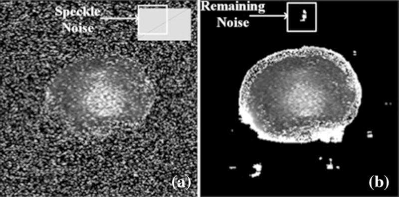
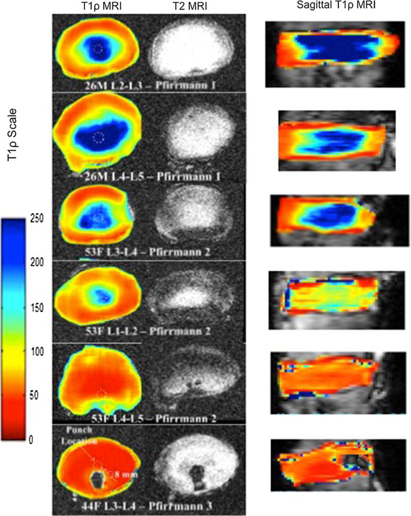
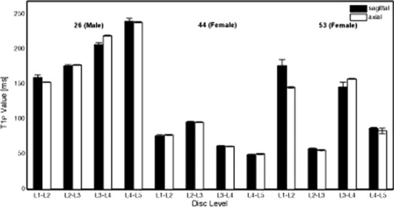
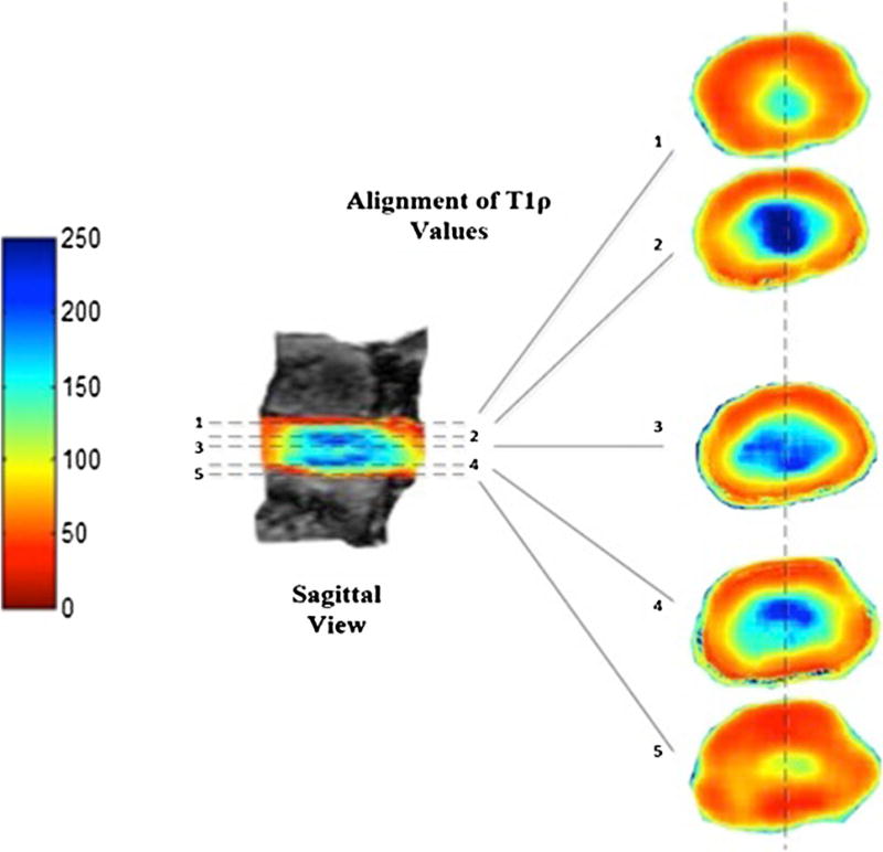
T1ρ and T2-weighted MR images of 12 intervertebral discs (IVDs) from 3 harvested human lumbar spines (levels L1-L2 to L5-S1) were grouped across their degenerative grade (Pfirrmann scores) and analyzed using a 3T MRI scanner in the axial and sagittal views. Post-processing of axial T1ρ-weighted images was performed using a Wiener filter. Median axial T1ρ values for traced regions of interest (ROIs) on color maps were compared against ROIs in the corresponding location in the sagittal plane of each disc. Assessment of sulfated glycosaminoglycans (GAGs) content was also performed.

RESULTS

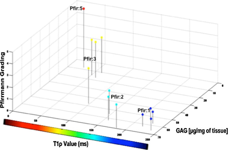
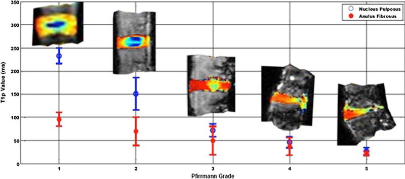
Comparison of post Wiener filtered mid-axial T1ρ values in the NP with corresponding mid-sagittal values revealed no statistical difference (P > 0.05). Higher axial T1ρ and biochemically measured GAGs content corresponded to a lower Pfirrmann grading of the IVDs. A strong association between the T1ρ values and the GAG contents was observed (r = 0.85, P = 0.0002).

CONCLUSIONS

The axial T1ρ methodology was validated against sagittal T1ρ providing an augmented spatial representation of IVD and can facilitate localization of focal degeneration within IVDs. T1ρ values provided a better granularity assessment of degenerative disc disease as it correlated with proteoglycan concentration. Thus, Wiener filtering is an effective tool for removing noise from T1ρ-weighted axial MR images.

摘要

目的

本研究旨在调查轴向T1ρ磁共振成像(MRI)在评估退变椎间盘(IDD)中蛋白聚糖浓度和含量方面是否与已确立的矢状面T1ρ MRI具有相似的准确性。

方法

从3具新鲜人腰椎(L1-L2至L5-S1节段)获取的12个椎间盘(IVD)的T1ρ和T2加权MR图像,根据其退变程度(Pfirrmann分级)分组,并使用3T MRI扫描仪在轴向和矢状面进行分析。轴向T1ρ加权图像的后处理采用维纳滤波器。将彩色图上感兴趣区域(ROI)的轴向T1ρ中位数与每个椎间盘矢状面相应位置的ROI进行比较。同时也对硫酸化糖胺聚糖(GAG)含量进行了评估。

结果

对经维纳滤波后的髓核轴向T1ρ中值与相应矢状面中值进行比较,未发现统计学差异(P>0.05)。较高的轴向T1ρ和生化测量的GAG含量对应较低的IVD Pfirrmann分级。观察到T1ρ值与GAG含量之间存在强相关性(r = 0.85,P = 0.0002)。

结论

轴向T1ρ方法经矢状面T1ρ验证有效,可提供IVD增强的空间图像,并有助于定位IVD内的局灶性退变。由于T1ρ值与蛋白聚糖浓度相关,因此能更好地对椎间盘退变疾病进行精细评估。因此,维纳滤波是去除T1ρ加权轴向MR图像噪声的有效工具。

相似文献

1
Axial T1ρ MRI as a diagnostic imaging modality to quantify proteoglycan concentration in degenerative disc disease.
Eur Spine J. 2015 Nov;24(11):2395-401. doi: 10.1007/s00586-014-3582-6. Epub 2014 Sep 19.
2
Glycosaminoglycan Chemical Exchange Saturation Transfer of Lumbar Intervertebral Discs in Healthy Volunteers.
Spine (Phila Pa 1976). 2016 Jan;41(2):146-52. doi: 10.1097/BRS.0000000000001144.
3
Assessment of human disc degeneration and proteoglycan content using T1rho-weighted magnetic resonance imaging.
Spine (Phila Pa 1976). 2006 May 15;31(11):1253-7. doi: 10.1097/01.brs.0000217708.54880.51.
4
T1ρ magnetic resonance imaging quantification of early lumbar intervertebral disc degeneration in healthy young adults.
Spine (Phila Pa 1976). 2012 Jun 15;37(14):1224-30. doi: 10.1097/BRS.0b013e31824b2450.
8
In vivo intervertebral disc mechanical deformation following a treadmill walking "stress test" is inversely related to T1rho relaxation time.
Osteoarthritis Cartilage. 2023 Jan;31(1):126-133. doi: 10.1016/j.joca.2022.09.008. Epub 2022 Sep 30.
10
Novel Application of the Pfirrmann Disc Degeneration Grading System to 9.4T MRI: Higher Reliability Compared to 3T MRI.
Spine (Phila Pa 1976). 2019 Jul 1;44(13):E766-E773. doi: 10.1097/BRS.0000000000002967.

引用本文的文献

3
Imaging of the spine: Where do we stand?
World J Radiol. 2019 Apr 28;11(4):55-61. doi: 10.4329/wjr.v11.i4.55.
8
Multimodal quantitative magnetic resonance imaging for lumbar intervertebral disc degeneration.
Exp Ther Med. 2017 Sep;14(3):2078-2084. doi: 10.3892/etm.2017.4786. Epub 2017 Jul 12.
9
T1ρ magnetic resonance: basic physics principles and applications in knee and intervertebral disc imaging.
Quant Imaging Med Surg. 2015 Dec;5(6):858-85. doi: 10.3978/j.issn.2223-4292.2015.12.06.

本文引用的文献

2
Biomechanics of disc degeneration.
Adv Orthop. 2012;2012:726210. doi: 10.1155/2012/726210. Epub 2012 Jun 17.
3
T1ρ magnetic resonance imaging quantification of early lumbar intervertebral disc degeneration in healthy young adults.
Spine (Phila Pa 1976). 2012 Jun 15;37(14):1224-30. doi: 10.1097/BRS.0b013e31824b2450.
4
Development of a bioreactor for axially loaded intervertebral disc organ culture.
Tissue Eng Part C Methods. 2011 Oct;17(10):1011-9. doi: 10.1089/ten.TEC.2011.0025. Epub 2011 Jul 19.
5
T1ρ magnetic resonance imaging and discography pressure as novel biomarkers for disc degeneration and low back pain.
Spine (Phila Pa 1976). 2011 Dec 1;36(25):2190-6. doi: 10.1097/BRS.0b013e31820287bf.
6
Validation of sodium magnetic resonance imaging of intervertebral disc.
Spine (Phila Pa 1976). 2010 Mar 1;35(5):505-10. doi: 10.1097/BRS.0b013e3181b32d3b.
7
Age-related changes in the extracellular matrix of nucleus pulposus and anulus fibrosus of human intervertebral disc.
Spine (Phila Pa 1976). 2009 Jan 1;34(1):10-6. doi: 10.1097/BRS.0b013e31818e5ddd.
8
Sequential anisotropic Wiener filtering applied to 3D MRI data.
Magn Reson Imaging. 2007 Feb;25(2):278-92. doi: 10.1016/j.mri.2006.05.001. Epub 2006 Dec 19.
9
Review of existing grading systems for cervical or lumbar disc and facet joint degeneration.
Eur Spine J. 2006 Jun;15(6):705-18. doi: 10.1007/s00586-005-0954-y. Epub 2005 Sep 20.

文献AI研究员

20分钟写一篇综述,助力文献阅读效率提升50倍。

立即体验

用中文搜PubMed

大模型驱动的PubMed中文搜索引擎

马上搜索

文档翻译

学术文献翻译模型,支持多种主流文档格式。

立即体验