Chow James C L
Radiation Medicine Program, Princess Margaret Cancer Centre, University Health Network, Toronto, ON M5G 1X6, Canada.
Department of Radiation Oncology, University of Toronto, Toronto, ON M5T 1P5, Canada.
Biomolecules. 2025 Mar 20;15(3):444. doi: 10.3390/biom15030444.
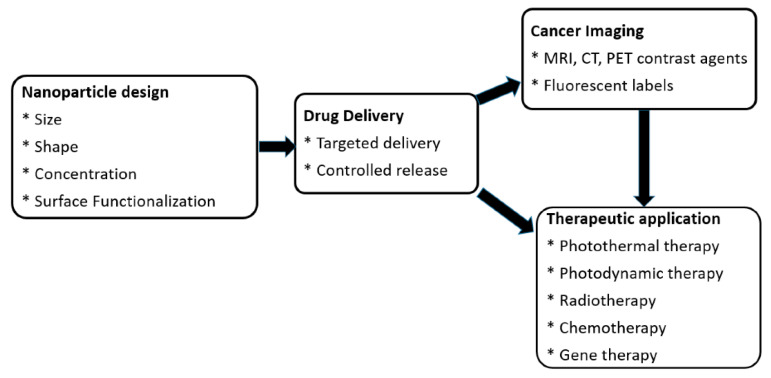
Nanomaterials represent an innovation in cancer imaging by offering enhanced contrast, improved targeting capabilities, and multifunctional imaging modalities. Recent advancements in material engineering have enabled the development of nanoparticles tailored for various imaging techniques, including magnetic resonance imaging (MRI), computed tomography (CT), positron emission tomography (PET), and ultrasound (US). These nanoscale agents improve sensitivity and specificity, enabling early cancer detection and precise tumor characterization. Monte Carlo (MC) simulations play a pivotal role in optimizing nanomaterial-based imaging by modeling their interactions with biological tissues, predicting contrast enhancement, and refining dosimetry for radiation-based imaging techniques. These computational methods provide valuable insights into nanoparticle behavior, aiding in the design of more effective imaging agents. Moreover, artificial intelligence (AI) and machine learning (ML) approaches are transforming cancer imaging by enhancing image reconstruction, automating segmentation, and improving diagnostic accuracy. AI-driven models can also optimize MC-based simulations by accelerating data analysis and refining nanoparticle design through predictive modeling. This review explores the latest advancements in nanomaterial-based cancer imaging, highlighting the synergy between nanotechnology, MC simulations, and AI-driven innovations. By integrating these interdisciplinary approaches, future cancer imaging technologies can achieve unprecedented precision, paving the way for more effective diagnostics and personalized treatment strategies.
纳米材料通过提供增强的对比度、改进的靶向能力和多功能成像方式,代表了癌症成像领域的一项创新。材料工程的最新进展使得能够开发出针对各种成像技术量身定制的纳米颗粒,包括磁共振成像(MRI)、计算机断层扫描(CT)、正电子发射断层扫描(PET)和超声(US)。这些纳米级试剂提高了灵敏度和特异性,能够实现癌症的早期检测和精确的肿瘤特征描述。蒙特卡罗(MC)模拟在优化基于纳米材料的成像中起着关键作用,它通过对纳米材料与生物组织的相互作用进行建模、预测对比度增强以及完善基于辐射的成像技术的剂量测定。这些计算方法为纳米颗粒的行为提供了有价值的见解,有助于设计更有效的成像试剂。此外,人工智能(AI)和机器学习(ML)方法正在通过增强图像重建、自动化分割和提高诊断准确性来改变癌症成像。人工智能驱动的模型还可以通过加速数据分析和通过预测建模完善纳米颗粒设计来优化基于MC的模拟。本综述探讨了基于纳米材料的癌症成像的最新进展,强调了纳米技术、MC模拟和人工智能驱动的创新之间的协同作用。通过整合这些跨学科方法,未来的癌症成像技术可以实现前所未有的精度,为更有效的诊断和个性化治疗策略铺平道路。