Hinamoto Norikazu, Maeshima Yohei, Yamasaki Hiroko, Nasu Tatsuyo, Saito Daisuke, Watatani Hiroyuki, Ujike Haruyo, Tanabe Katsuyuki, Masuda Kana, Arata Yuka, Sugiyama Hitoshi, Sato Yasufumi, Makino Hirofumi
Department of Medicine and Clinical Science, Okayama University Graduate School of Medicine, Dentistry and Pharmaceutical Sciences, Okayama, Japan.
Department of Chronic Kidney Disease and Cardiovascular Disease, Okayama University Graduate School of Medicine, Dentistry and Pharmaceutical Sciences, Okayama, Japan.
PLoS One. 2014 Sep 25;9(9):e107934. doi: 10.1371/journal.pone.0107934. eCollection 2014.
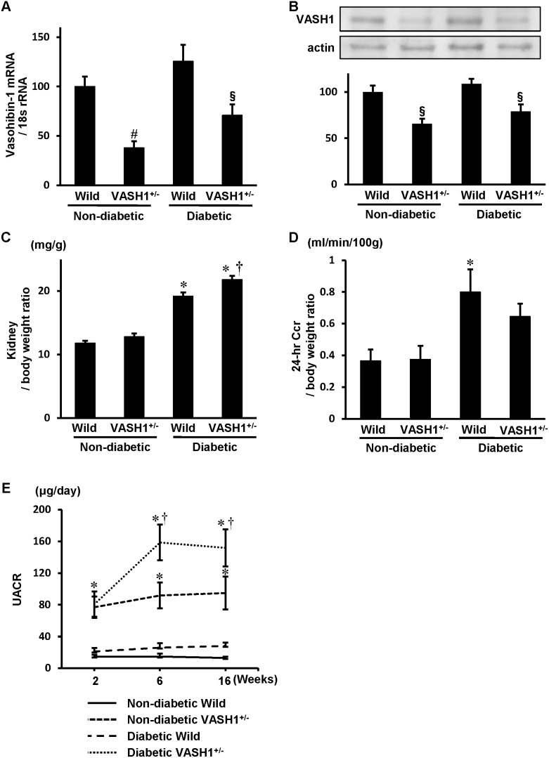
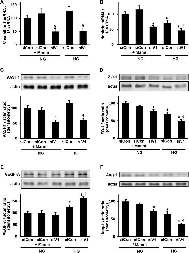
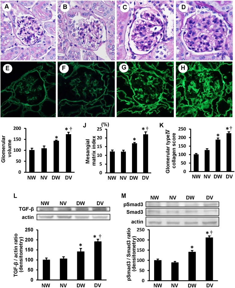
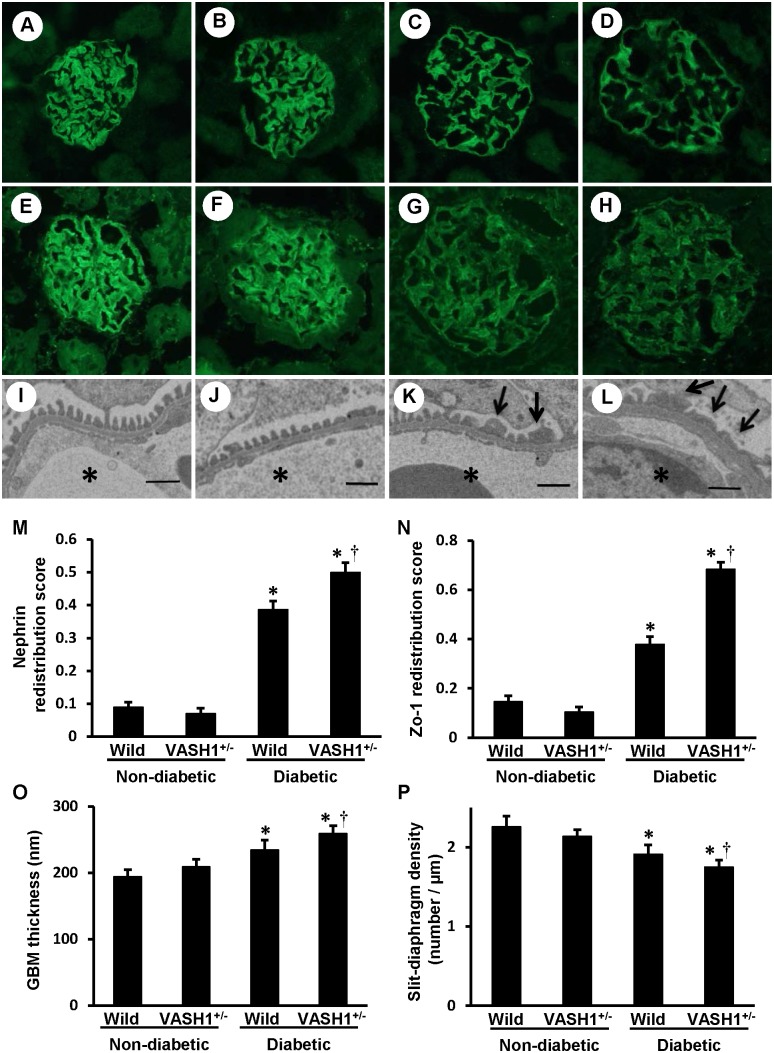
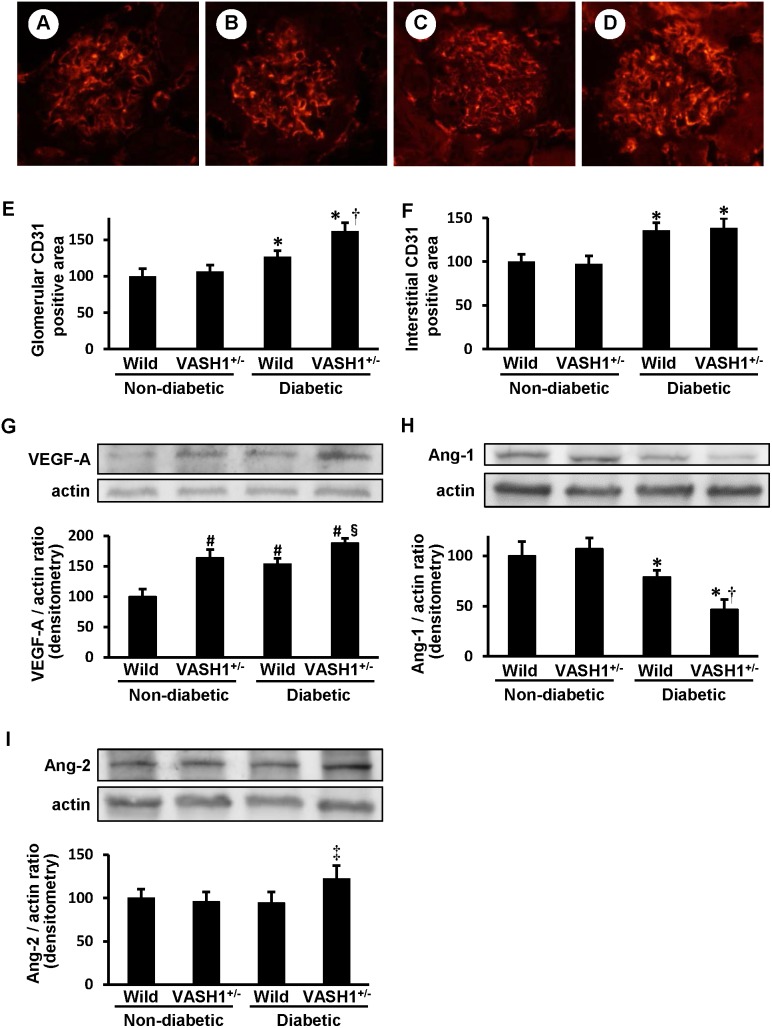
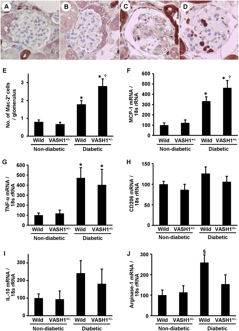
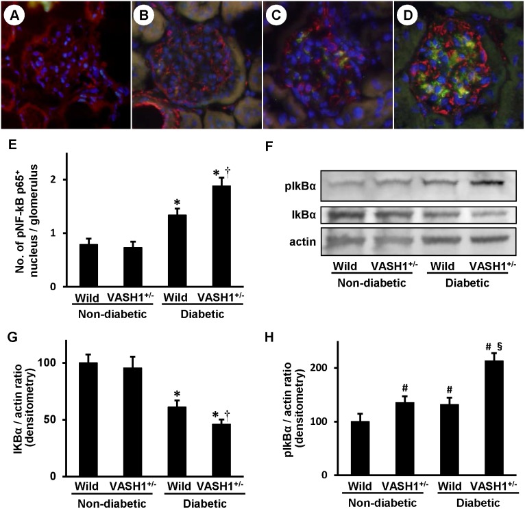
Vasohibin-1 (VASH1) is a unique endogenous inhibitor of angiogenesis that is induced in endothelial cells by pro-angiogenic factors. We previously reported renoprotective effect of adenoviral delivery of VASH1 in diabetic nephropathy model, and herein investigated the potential protective role of endogenous VASH1 by using VASH1-deficient mice. Streptozotocin-induced type 1 diabetic VASH1 heterozygous knockout mice (VASH1(+/-)) or wild-type diabetic mice were sacrificed 16 weeks after inducing diabetes. In the diabetic VASH1(+/-) mice, albuminuria were significantly exacerbated compared with the diabetic wild-type littermates, in association with the dysregulated distribution of glomerular slit diaphragm related proteins, nephrin and ZO-1, glomerular basement membrane thickening and reduction of slit diaphragm density. Glomerular monocyte/macrophage infiltration and glomerular nuclear translocation of phosphorylated NF-κB p65 were significantly exacerbated in the diabetic VASH1(+/-) mice compared with the diabetic wild-type littermates, accompanied by the augmentation of VEGF-A, M1 macrophage-derived MCP-1 and phosphorylation of IκBα, and the decrease of angiopoietin-1/2 ratio and M2 macrophage-derived Arginase-1. The glomerular CD31(+) endothelial area was also increased in the diabetic VASH1(+/-) mice compared with the diabetic-wild type littermates. Furthermore, the renal and glomerular hypertrophy, glomerular accumulation of mesangial matrix and type IV collagen and activation of renal TGF-β1/Smad3 signaling, a key mediator of renal fibrosis, were exacerbated in the diabetic VASH1(+/-) mice compared with the diabetic wild-type littermates. In conditionally immortalized mouse podocytes cultured under high glucose condition, transfection of VASH1 small interfering RNA (siRNA) resulted in the reduction of nephrin, angiopoietin-1 and ZO-1, and the augmentation of VEGF-A compared with control siRNA. These results suggest that endogenous VASH1 may regulate the development of diabetic renal alterations, partly via direct effects on podocytes, and thus, a strategy to recover VASH1 might potentially lead to the development of a novel therapeutic approach for diabetic nephropathy.
血管抑制素-1(VASH1)是一种独特的内源性血管生成抑制剂,由促血管生成因子在内皮细胞中诱导产生。我们之前报道了腺病毒介导的VASH1在糖尿病肾病模型中的肾脏保护作用,在此我们使用VASH1基因缺陷小鼠研究内源性VASH1的潜在保护作用。链脲佐菌素诱导的1型糖尿病VASH1杂合敲除小鼠(VASH1(+/-))或野生型糖尿病小鼠在诱导糖尿病16周后处死。在糖尿病VASH1(+/-)小鼠中,与糖尿病野生型同窝小鼠相比,蛋白尿显著加重,同时伴有肾小球裂孔隔膜相关蛋白、nephrin和ZO-1的分布失调、肾小球基底膜增厚以及裂孔隔膜密度降低。与糖尿病野生型同窝小鼠相比,糖尿病VASH1(+/-)小鼠肾小球单核细胞/巨噬细胞浸润和磷酸化NF-κB p65的肾小球核转位显著加重,同时伴有VEGF-A、M1巨噬细胞衍生的MCP-1增加以及IκBα磷酸化,血管生成素-1/2比值和M2巨噬细胞衍生的精氨酸酶-1降低。与糖尿病野生型同窝小鼠相比,糖尿病VASH1(+/-)小鼠肾小球CD31(+)内皮面积也增加。此外,与糖尿病野生型同窝小鼠相比,糖尿病VASH1(+/-)小鼠的肾脏和肾小球肥大、系膜基质和IV型胶原在肾小球的积聚以及肾纤维化的关键介质肾TGF-β1/Smad3信号通路的激活均加重。在高糖条件下培养的条件性永生化小鼠足细胞中,与对照小干扰RNA相比,转染VASH1小干扰RNA(siRNA)导致nephrin、血管生成素-1和ZO-1减少,VEGF-A增加。这些结果表明,内源性VASH1可能部分通过对足细胞的直接作用来调节糖尿病肾脏病变的发展,因此,恢复VASH1的策略可能会潜在地导致开发一种新的糖尿病肾病治疗方法。