• 文献检索
  • 文档翻译
  • 深度研究
  • 学术资讯
  • Suppr Zotero 插件Zotero 插件
  • 邀请有礼
  • 套餐&价格
  • 历史记录
应用&插件
Suppr Zotero 插件Zotero 插件浏览器插件Mac 客户端Windows 客户端微信小程序
定价
高级版会员购买积分包购买API积分包
服务
文献检索文档翻译深度研究API 文档MCP 服务
关于我们
关于 Suppr公司介绍联系我们用户协议隐私条款
关注我们

Suppr 超能文献

核心技术专利:CN118964589B侵权必究
粤ICP备2023148730 号-1Suppr @ 2026

文献检索

告别复杂PubMed语法,用中文像聊天一样搜索,搜遍4000万医学文献。AI智能推荐,让科研检索更轻松。

立即免费搜索

文件翻译

保留排版,准确专业,支持PDF/Word/PPT等文件格式,支持 12+语言互译。

免费翻译文档

深度研究

AI帮你快速写综述,25分钟生成高质量综述,智能提取关键信息,辅助科研写作。

立即免费体验

视紫红质在天然人类和小鼠视杆外段盘膜中的纳米结构组织。

Nanodomain organization of rhodopsin in native human and murine rod outer segment disc membranes.

作者信息

Whited Allison M, Park Paul S-H

机构信息

Department of Ophthalmology and Visual Sciences, Case Western Reserve University, Cleveland, OH 44106, USA.

Department of Ophthalmology and Visual Sciences, Case Western Reserve University, Cleveland, OH 44106, USA.

出版信息

Biochim Biophys Acta. 2015 Jan;1848(1 Pt A):26-34. doi: 10.1016/j.bbamem.2014.10.007. Epub 2014 Oct 12.

DOI:10.1016/j.bbamem.2014.10.007
PMID:25305340
原文链接:https://pmc.ncbi.nlm.nih.gov/articles/PMC4259819/
Abstract

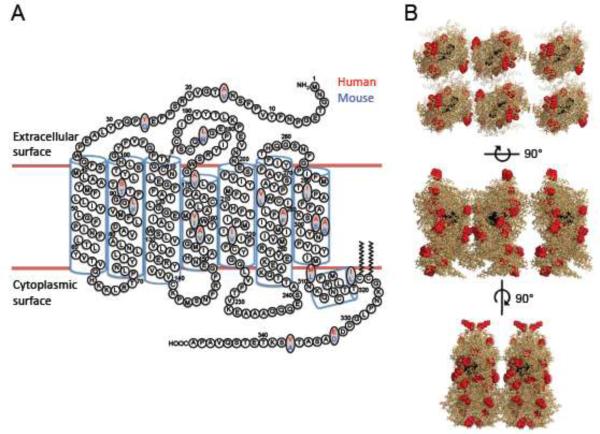
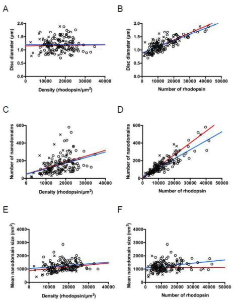
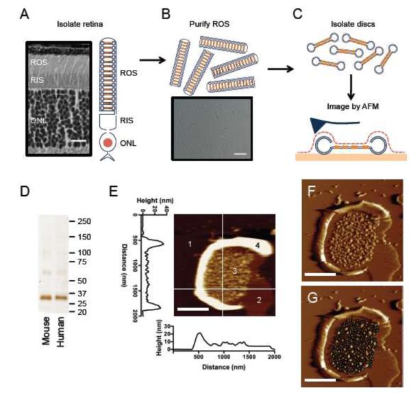
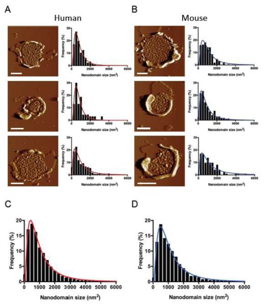
Biological membranes display distinct domains that organize membrane proteins and signaling molecules to facilitate efficient and reliable signaling. The organization of rhodopsin, a G protein-coupled receptor, in native rod outer segment disc membranes was investigated by atomic force microscopy. Atomic force microscopy revealed that rhodopsin is arranged into domains of variable size, which we refer to herein as nanodomains, in native membranes. Quantitative analysis of 150 disc membranes revealed that the physical properties of nanodomains are conserved in humans and mice and that the properties of individual disc membranes can be variable. Examining the variable properties of disc membranes revealed some of the factors contributing to the size of rod outer segment discs and the formation of nanodomains in the membrane. The diameter of rod outer segment discs was dependent on the number of rhodopsin molecules incorporated into the membrane but independent of the spatial density of rhodopsin. The number of nanodomains present in a single disc was also dependent on the number of rhodopsin molecules incorporated into the membrane. The size of the nanodomains was largely independent of the number or spatial density of rhodopsin in the membrane.

摘要

生物膜呈现出不同的结构域,这些结构域可组织膜蛋白和信号分子,以促进高效且可靠的信号传导。利用原子力显微镜研究了视紫红质(一种G蛋白偶联受体)在天然视杆细胞外段盘状膜中的组织方式。原子力显微镜显示,在天然膜中视紫红质排列成大小可变的结构域,在此我们将其称为纳米结构域。对150个盘状膜进行的定量分析表明,纳米结构域的物理特性在人类和小鼠中是保守的,并且单个盘状膜的特性可能存在差异。研究盘状膜的可变特性揭示了一些影响视杆细胞外段盘大小以及膜中纳米结构域形成的因素。视杆细胞外段盘的直径取决于掺入膜中的视紫红质分子数量,但与视紫红质的空间密度无关。单个盘中存在的纳米结构域数量也取决于掺入膜中的视紫红质分子数量。纳米结构域的大小在很大程度上与膜中视紫红质的数量或空间密度无关。

https://cdn.ncbi.nlm.nih.gov/pmc/blobs/71d6/4259819/4079e6f6654f/nihms-634256-f0004.jpg
https://cdn.ncbi.nlm.nih.gov/pmc/blobs/71d6/4259819/3242b50034c7/nihms-634256-f0001.jpg
https://cdn.ncbi.nlm.nih.gov/pmc/blobs/71d6/4259819/dc3637e40297/nihms-634256-f0002.jpg
https://cdn.ncbi.nlm.nih.gov/pmc/blobs/71d6/4259819/f5b7b83060f0/nihms-634256-f0003.jpg
https://cdn.ncbi.nlm.nih.gov/pmc/blobs/71d6/4259819/4079e6f6654f/nihms-634256-f0004.jpg

相似文献

1
Nanodomain organization of rhodopsin in native human and murine rod outer segment disc membranes.视紫红质在天然人类和小鼠视杆外段盘膜中的纳米结构组织。
Biochim Biophys Acta. 2015 Jan;1848(1 Pt A):26-34. doi: 10.1016/j.bbamem.2014.10.007. Epub 2014 Oct 12.
2
Rhodopsin Forms Nanodomains in Rod Outer Segment Disc Membranes of the Cold-Blooded Xenopus laevis.视紫红质在冷血动物非洲爪蟾的视杆外段盘膜中形成纳米结构域。
PLoS One. 2015 Oct 22;10(10):e0141114. doi: 10.1371/journal.pone.0141114. eCollection 2015.
3
Investigating the Nanodomain Organization of Rhodopsin in Native Membranes by Atomic Force Microscopy.利用原子力显微镜研究天然膜中视紫红质的纳米域组织。
Methods Mol Biol. 2019;1886:61-74. doi: 10.1007/978-1-4939-8894-5_4.
4
Impact of reduced rhodopsin expression on the structure of rod outer segment disc membranes.视紫红质表达降低对视杆细胞外节盘膜结构的影响。
Biochemistry. 2015 May 12;54(18):2885-94. doi: 10.1021/acs.biochem.5b00003. Epub 2015 Apr 27.
5
Adaptations in rod outer segment disc membranes in response to environmental lighting conditions.对环境光照条件的 rods 外段盘膜的适应。
Biochim Biophys Acta Mol Cell Res. 2017 Oct;1864(10):1691-1702. doi: 10.1016/j.bbamcr.2017.06.013. Epub 2017 Jun 20.
6
The supramolecular structure of the GPCR rhodopsin in solution and native disc membranes.溶液中和天然盘状膜中G蛋白偶联受体视紫红质的超分子结构。
Mol Membr Biol. 2004 Nov-Dec;21(6):435-46. doi: 10.1080/09687860400020291.
7
Raftophilic rhodopsin-clusters offer stochastic platforms for G protein signalling in retinal discs.筏筏亲视网膜紫质簇为视网膜盘上的 G 蛋白信号提供了随机平台。
Commun Biol. 2019 Jun 14;2:209. doi: 10.1038/s42003-019-0459-6. eCollection 2019.
8
Rhodopsin signaling and organization in heterozygote rhodopsin knockout mice.杂合子视紫红质基因敲除小鼠中的视紫红质信号传导与组织
J Biol Chem. 2004 Nov 12;279(46):48189-96. doi: 10.1074/jbc.M408362200. Epub 2004 Aug 26.
9
Rhodopsin is spatially heterogeneously distributed in rod outer segment disk membranes.视紫红质在杆状细胞外段盘膜中呈空间异质分布。
J Mol Recognit. 2011 May-Jun;24(3):483-9. doi: 10.1002/jmr.1086.
10
The G protein-coupled receptor rhodopsin in the native membrane.天然膜中的G蛋白偶联受体视紫红质。
FEBS Lett. 2004 Apr 30;564(3):281-288. doi: 10.1016/S0014-5793(04)00194-2.

引用本文的文献

1
Understanding the Rhodopsin Worldview Through Atomic Force Microscopy (AFM): Structure, Stability, and Activity Studies.通过原子力显微镜(AFM)了解视紫红质的世界观:结构、稳定性和活性研究。
Chem Rec. 2023 Oct;23(10):e202300113. doi: 10.1002/tcr.202300113. Epub 2023 Jun 2.
2
Kinetics of cone specific G-protein signaling in avian photoreceptor cells.禽类光感受器细胞中视锥特异性G蛋白信号转导的动力学
Front Mol Neurosci. 2023 Jan 17;16:1107025. doi: 10.3389/fnmol.2023.1107025. eCollection 2023.
3
A hybrid stochastic/deterministic model of single photon response and light adaptation in mouse rods.

本文引用的文献

1
Explicit spatiotemporal simulation of receptor-G protein coupling in rod cell disk membranes.视杆细胞盘膜中受体 - G 蛋白偶联的显式时空模拟。
Biophys J. 2014 Sep 2;107(5):1042-1053. doi: 10.1016/j.bpj.2014.05.050.
2
G protein-coupled receptor oligomerization revisited: functional and pharmacological perspectives.G蛋白偶联受体寡聚化再探讨:功能与药理学视角
Pharmacol Rev. 2014 Feb 10;66(2):413-34. doi: 10.1124/pr.113.008052. Print 2014.
3
Regulation of rhodopsin-eGFP distribution in transgenic xenopus rod outer segments by light.
小鼠视杆细胞中单光子响应和光适应的混合随机/确定性模型。
Comput Struct Biotechnol J. 2021 Jun 23;19:3720-3734. doi: 10.1016/j.csbj.2021.06.033. eCollection 2021.
4
Supramolecular organization of rhodopsin in rod photoreceptor cell membranes.视紫红质在视杆感光细胞膜中的超分子组织。
Pflugers Arch. 2021 Sep;473(9):1361-1376. doi: 10.1007/s00424-021-02522-5. Epub 2021 Feb 16.
5
Differential Aggregation Properties of Mutant Human and Bovine Rhodopsin.突变型人源和牛视紫红质的差异聚集特性。
Biochemistry. 2021 Jan 12;60(1):6-18. doi: 10.1021/acs.biochem.0c00733. Epub 2020 Dec 27.
6
Loss of PRCD alters number and packaging density of rhodopsin in rod photoreceptor disc membranes.PRCD 缺失改变了视杆感光细胞盘膜中视紫红质的数量和包装密度。
Sci Rep. 2020 Oct 21;10(1):17885. doi: 10.1038/s41598-020-74628-2.
7
Differential adaptations in rod outer segment disc membranes in different models of congenital stationary night blindness.不同先天性静止性夜盲症模型中视杆外段盘膜的差异适应性。
Biochim Biophys Acta Biomembr. 2020 Oct 1;1862(10):183396. doi: 10.1016/j.bbamem.2020.183396. Epub 2020 Jun 11.
8
Affinity of rhodopsin to raft enables the aligned oligomer formation from dimers: Coarse-grained molecular dynamics simulation of disk membranes.视紫红质与筏的亲和力使二聚体从对齐的寡聚体形成:盘膜的粗粒度分子动力学模拟。
PLoS One. 2020 Feb 7;15(2):e0226123. doi: 10.1371/journal.pone.0226123. eCollection 2020.
9
Rhodopsin Oligomerization and Aggregation.视紫红质寡聚化和聚集。
J Membr Biol. 2019 Oct;252(4-5):413-423. doi: 10.1007/s00232-019-00078-1. Epub 2019 Jul 8.
10
Raftophilic rhodopsin-clusters offer stochastic platforms for G protein signalling in retinal discs.筏筏亲视网膜紫质簇为视网膜盘上的 G 蛋白信号提供了随机平台。
Commun Biol. 2019 Jun 14;2:209. doi: 10.1038/s42003-019-0459-6. eCollection 2019.
光照调控转基因非洲爪蟾视杆外节细胞中视蛋白-eGFP 的分布
PLoS One. 2013 Nov 15;8(11):e80059. doi: 10.1371/journal.pone.0080059. eCollection 2013.
4
Membrane driven spatial organization of GPCRs.G蛋白偶联受体的膜驱动空间组织
Sci Rep. 2013 Oct 9;3:2909. doi: 10.1038/srep02909.
5
Nanoscale structural features determined by AFM for single virus particles.原子力显微镜测定的单个病毒颗粒的纳米结构特征。
Nanoscale. 2013 Nov 21;5(22):10877-86. doi: 10.1039/c3nr02706f. Epub 2013 Sep 9.
6
A physiological role for the supramolecular organization of rhodopsin and transducin in rod photoreceptors.视紫红质和转导蛋白的超分子组织在杆状光感受器中的生理作用。
FEBS Lett. 2013 Jun 27;587(13):2060-6. doi: 10.1016/j.febslet.2013.05.017. Epub 2013 May 16.
7
Atomic force microscopy: a multifaceted tool to study membrane proteins and their interactions with ligands.原子力显微镜:一种用于研究膜蛋白及其与配体相互作用的多面工具。
Biochim Biophys Acta. 2014 Jan;1838(1 Pt A):56-68. doi: 10.1016/j.bbamem.2013.04.011. Epub 2013 Apr 16.
8
Spatial partitioning improves the reliability of biochemical signaling.空间分隔提高了生化信号传递的可靠性。
Proc Natl Acad Sci U S A. 2013 Apr 9;110(15):5927-32. doi: 10.1073/pnas.1218301110. Epub 2013 Mar 25.
9
The rhodopsin-transducin complex houses two distinct rhodopsin molecules.视紫红质转导复合物包含两个不同的视紫红质分子。
J Struct Biol. 2013 May;182(2):164-72. doi: 10.1016/j.jsb.2013.02.014. Epub 2013 Feb 28.
10
Asymmetry of the rhodopsin dimer in complex with transducin.视紫红质二聚体与转导蛋白复合物的非对称性。
FASEB J. 2013 Apr;27(4):1572-84. doi: 10.1096/fj.12-225383. Epub 2013 Jan 9.