Wheeler S E, Clark A M, Taylor D P, Young C L, Pillai V C, Stolz D B, Venkataramanan R, Lauffenburger D, Griffith L, Wells A
Department of Pathology, University of Pittsburgh, S711 Scaife Hall, 3550 Terrace Street, Pittsburgh, PA, USA.
1] Department of Pathology, University of Pittsburgh, S711 Scaife Hall, 3550 Terrace Street, Pittsburgh, PA, USA [2] Department of Bioengineering, University of Pittsburgh, Pittsburgh, PA, USA.
Br J Cancer. 2014 Dec 9;111(12):2342-50. doi: 10.1038/bjc.2014.533. Epub 2014 Oct 14.
Metastatic outgrowth in breast cancer can occur years after a seeming cure. Existing model systems of dormancy are limited as they do not recapitulate human metastatic dormancy without exogenous manipulations and are unable to query early events of micrometastases.
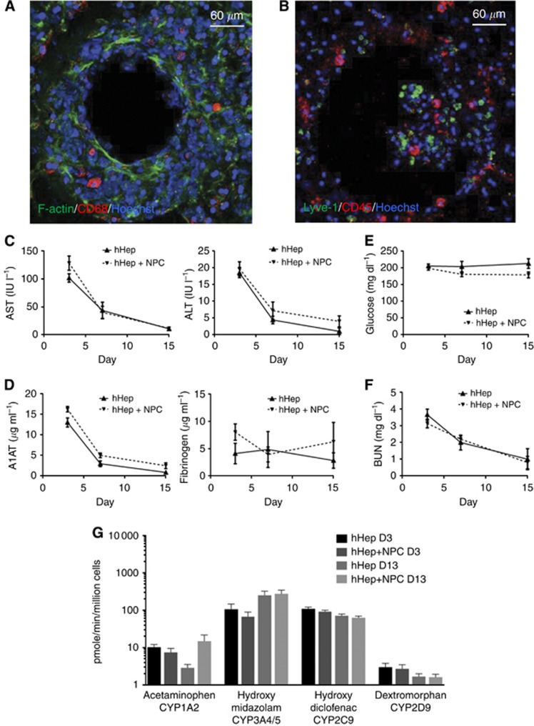
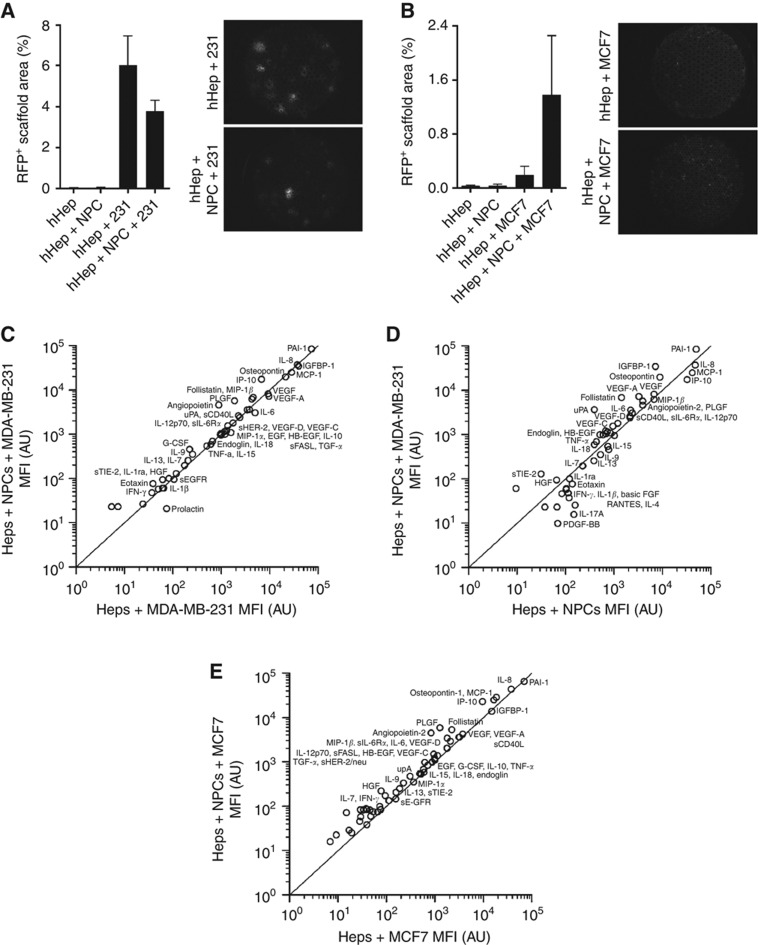
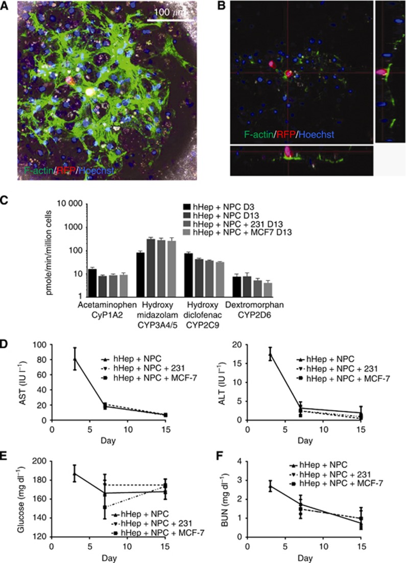
Here, we describe a human ex vivo hepatic microphysiologic system. The system is established with fresh human hepatocytes and non-parenchymal cells (NPCs) creating a microenvironment into which breast cancer cells (MCF7 and MDA-MB-231) are added.
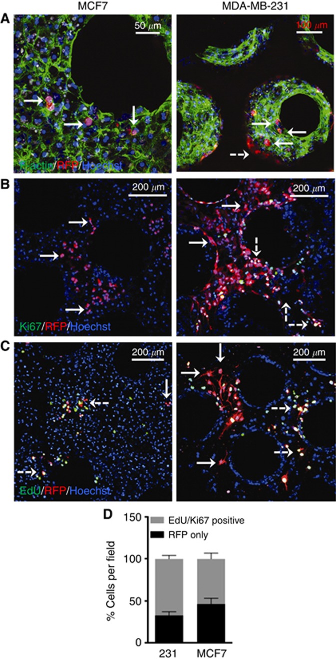
The hepatic tissue maintains function through 15 days as verified by liver-specific protein production and drug metabolism assays. The NPCs form an integral part of the hepatic niche, demonstrated within the system through their participation in differential signalling cascades and cancer cell outcomes. Breast cancer cells intercalate into the hepatic niche without interfering with hepatocyte function. Examination of cancer cells demonstrated that a significant subset enter a quiescent state of dormancy as shown by lack of cell cycling (EdU(-) or Ki67(-)). The presence of NPCs altered the cancer cell fraction entering quiescence, and lead to differential cytokine profiles in the microenvironment effluent.
These findings establish the liver microphysiologic system as a relevant model for the study of breast cancer metastases and entry into dormancy.
乳腺癌转移灶的生长可能在看似治愈数年之后出现。现有的休眠模型系统存在局限性,因为在没有外源干预的情况下,它们无法模拟人类转移性休眠,并且无法探究微转移的早期事件。
在此,我们描述了一种人源离体肝脏微生理系统。该系统由新鲜人肝细胞和非实质细胞(NPC)构建而成,形成一个微环境,然后将乳腺癌细胞(MCF7和MDA-MB-231)添加到其中。
通过肝脏特异性蛋白生成和药物代谢分析验证,肝脏组织在15天内保持功能。NPC构成肝脏生态位的一个组成部分,在系统中通过它们参与不同的信号级联反应和癌细胞结局得以证明。乳腺癌细胞嵌入肝脏生态位而不干扰肝细胞功能。对癌细胞的检测表明,相当一部分癌细胞进入静止休眠状态,表现为缺乏细胞周期(EdU阴性或Ki67阴性)。NPC的存在改变了进入静止状态的癌细胞比例,并导致微环境流出物中细胞因子谱的差异。
这些发现确立了肝脏微生理系统作为研究乳腺癌转移和进入休眠的相关模型。