Kamimura Mikio, Nakamura Yukio, Uchiyama Shigeharu, Ikegami Shota, Mukaiyama Keijiro, Kato Hiroyuki
Center of Osteoporosis and Spinal Disorders: Kamimura Orthopaedic Clinic, Matsumoto 399-0021, Japan.
Department of Orthopaedic Surgery, Shinshu University School of Medicine, Asahi 3-1-1, Matsumoto 390-8621, Japan.
Open Rheumatol J. 2014 Oct 2;8:46-53. doi: 10.2174/1874312901408010046. eCollection 2014.
This study examined hip osteoarthritis (OA) patients with joint pain and accompanying signal changes detected by magnetic resonance imaging (MRI).
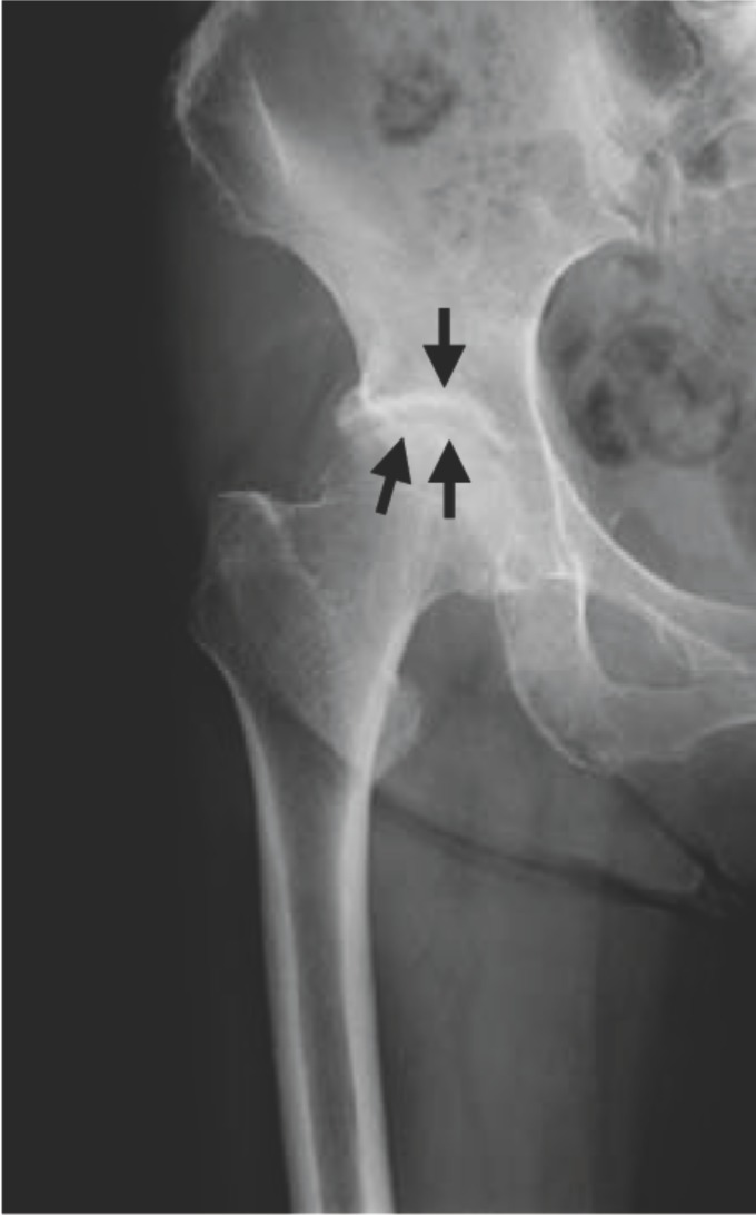
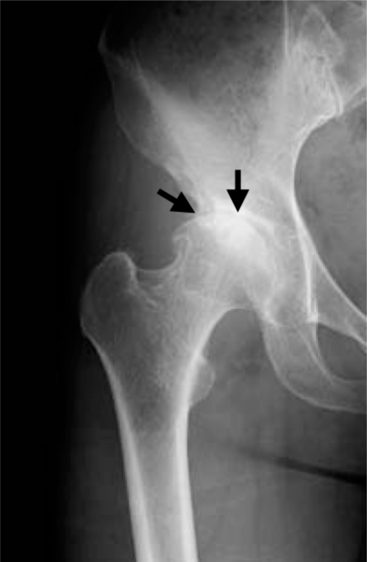
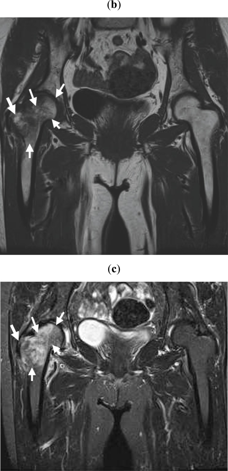
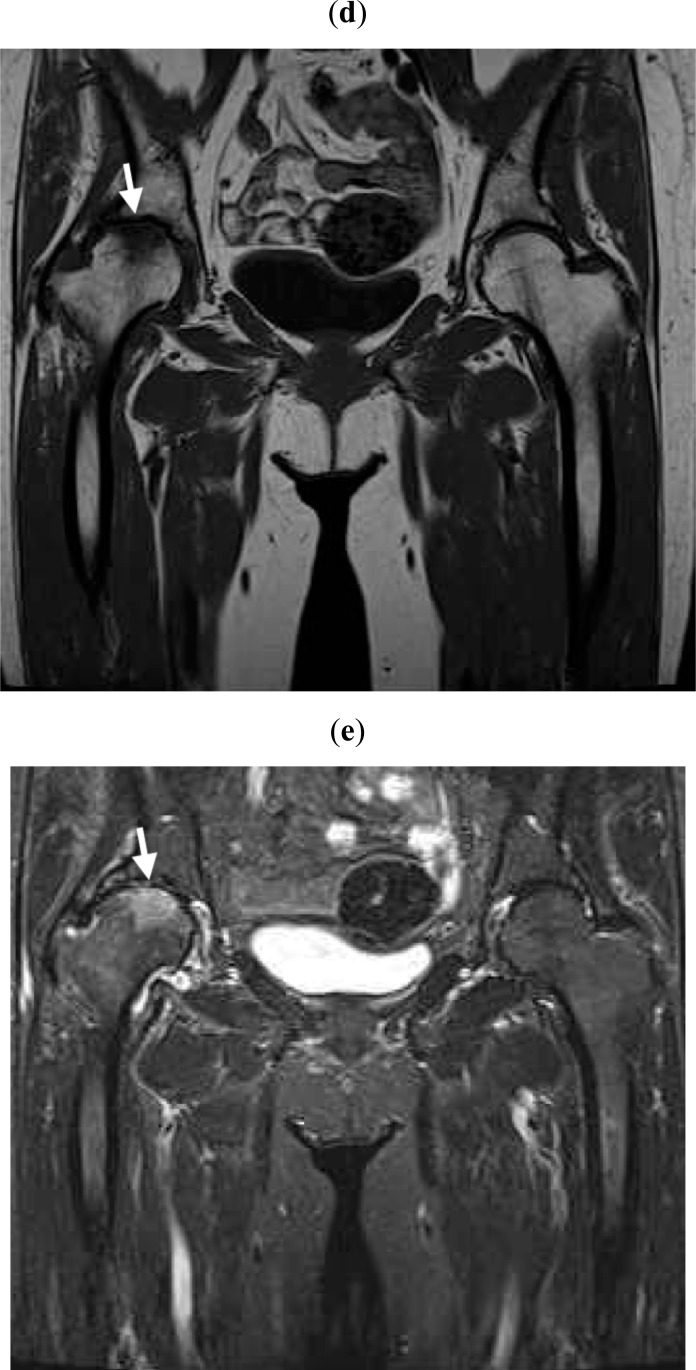
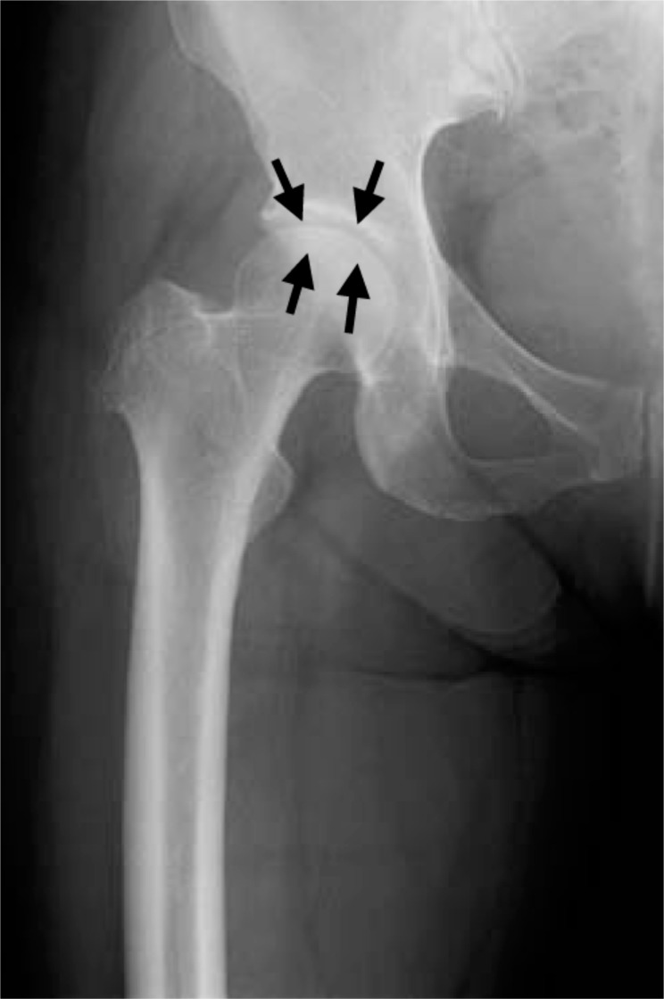
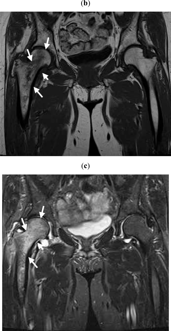
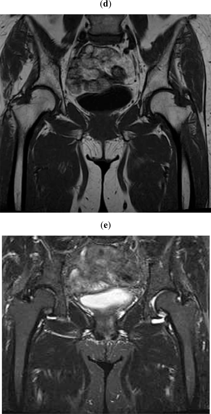
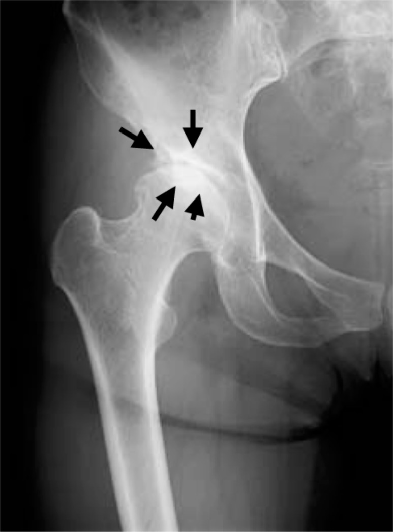
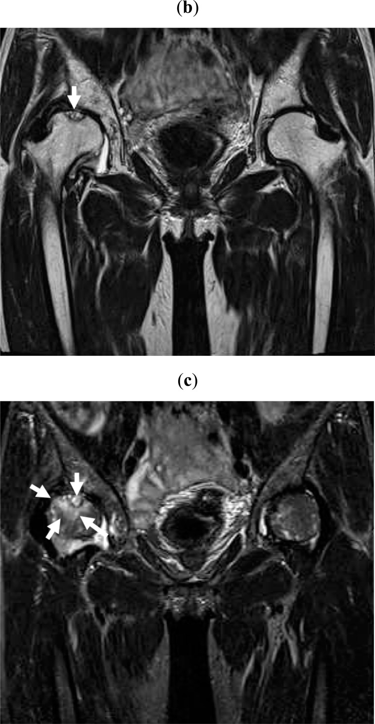
A total of 19 hip OA patients with suddenly occurring or worsening pain regardless of Kellgren-Lawrence grading were enrolled. The patients were monitored using MRI, plain radiographs, and the Denis pain scale for a minimum of 6 months. The patients were classified into 2 groups: those whose pain improved during conservative treatment (Group A) and those whose pain persisted (Group B).
Joint pain disappeared or was markedly improved in all 10 cases in Group A. Radiographic OA progression occurred in 7 of 8 cases with available radiographs. Hip MRI was performed on 7 of 10 patients, among whom bone signal changes disappeared in 6 patients. One patient exhibited persisting bone signal alterations although joint pain had completely disappeared. In Group B, joint pain remained in all 9 cases. Radiographic OA progression occurred in 8 of 9 cases, and local (4 cases) or broad (5 cases) bone signal alterations were present in end-point MRI examinations. Two patients exhibited different regional MRI bone signal changes (local or broad) at the end of follow-up. The mean age of Group B was significantly higher than that of Group A.
THIS STUDY UNCOVERED THE FOLLOWING OBSERVATIONS: 1) hip OA with joint pain had bone alterations that were detectable by MRI, 2) these bone alterations disappeared when joint pain improved, 3) bone alterations remained when joint pain continued, and 4) radiographic OA progressed to a more advanced stage over a short time period. These findings indicate that the pathophysiology of OA, joint pain, and OA progression may primarily be due to bone changes.
本研究对患有关节疼痛且磁共振成像(MRI)检测到伴有信号改变的髋骨关节炎(OA)患者进行了检查。
共纳入19例髋OA患者,这些患者不论Kellgren-Lawrence分级如何,均出现突然发作或加重的疼痛。使用MRI、X线平片和丹尼斯疼痛量表对患者进行至少6个月的监测。患者被分为两组:保守治疗期间疼痛改善的患者(A组)和疼痛持续的患者(B组)。
A组的所有10例患者关节疼痛消失或明显改善。8例有可用X线片的患者中,7例出现影像学OA进展。10例患者中的7例进行了髋部MRI检查,其中6例患者的骨信号改变消失。1例患者尽管关节疼痛已完全消失,但骨信号改变持续存在。B组的所有9例患者关节疼痛均持续。9例患者中的8例出现影像学OA进展,终点MRI检查显示局部(4例)或广泛(5例)骨信号改变。2例患者在随访结束时出现不同区域的MRI骨信号改变(局部或广泛)。B组的平均年龄显著高于A组。
本研究发现了以下情况:1)伴有关节疼痛的髋OA存在MRI可检测到的骨改变;2)关节疼痛改善时,这些骨改变消失;3)关节疼痛持续时,骨改变持续存在;4)影像学OA在短时间内进展到更晚期阶段。这些发现表明,OA的病理生理学、关节疼痛和OA进展可能主要归因于骨改变。