• 文献检索
  • 文档翻译
  • 深度研究
  • 学术资讯
  • Suppr Zotero 插件Zotero 插件
  • 邀请有礼
  • 套餐&价格
  • 历史记录
应用&插件
Suppr Zotero 插件Zotero 插件浏览器插件Mac 客户端Windows 客户端微信小程序
定价
高级版会员购买积分包购买API积分包
服务
文献检索文档翻译深度研究API 文档MCP 服务
关于我们
关于 Suppr公司介绍联系我们用户协议隐私条款
关注我们

Suppr 超能文献

核心技术专利:CN118964589B侵权必究
粤ICP备2023148730 号-1Suppr @ 2026

文献检索

告别复杂PubMed语法,用中文像聊天一样搜索,搜遍4000万医学文献。AI智能推荐,让科研检索更轻松。

立即免费搜索

文件翻译

保留排版,准确专业,支持PDF/Word/PPT等文件格式,支持 12+语言互译。

免费翻译文档

深度研究

AI帮你快速写综述,25分钟生成高质量综述,智能提取关键信息,辅助科研写作。

立即免费体验

使用共光路衍射光学层析成像技术对单个人类红细胞进行分析。

Profiling individual human red blood cells using common-path diffraction optical tomography.

作者信息

Kim Youngchan, Shim Hyoeun, Kim Kyoohyun, Park HyunJoo, Jang Seongsoo, Park YongKeun

机构信息

Department of Physics, Korea Advanced Institute of Science and Technology, Daejeon 305-701, Republic of Korea.

Department of Laboratory Medicine, University of Ulsan, College of Medicine and Asan Medical Center, Seoul 138-736, Republic of Korea.

出版信息

Sci Rep. 2014 Oct 17;4:6659. doi: 10.1038/srep06659.

DOI:10.1038/srep06659
PMID:25322756
原文链接:https://pmc.ncbi.nlm.nih.gov/articles/PMC4200412/
Abstract

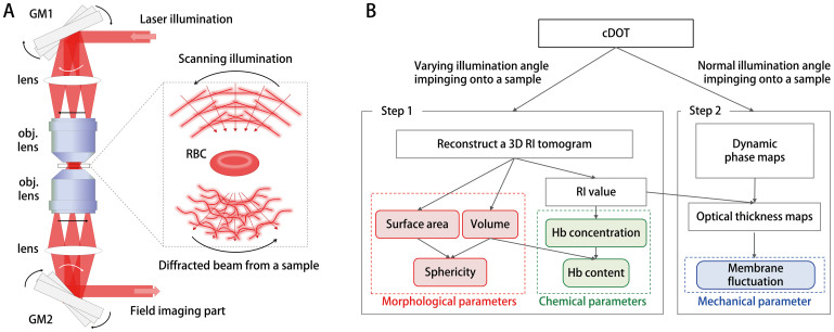
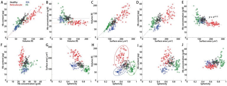
Due to its strong correlation with the pathophysiology of many diseases, information about human red blood cells (RBCs) has a crucial function in hematology. Therefore, measuring and understanding the morphological, chemical, and mechanical properties of individual RBCs is a key to understanding the pathophysiology of a number of diseases in hematology, as well as to opening up new possibilities for diagnosing diseases in their early stages. In this study, we present the simultaneous and quantitative measurement of the morphological, chemical, and mechanical parameters of individual RBCs employing optical holographic microtomography. In addition, it is demonstrated that the correlation analyses of these RBC parameters provide unique information for distinguishing and understanding diseases.

摘要

由于其与多种疾病的病理生理学密切相关,有关人类红细胞(RBC)的信息在血液学中具有至关重要的作用。因此,测量和了解单个红细胞的形态、化学和力学特性是理解血液学中多种疾病病理生理学的关键,也是为疾病早期诊断开辟新可能性的关键。在本研究中,我们展示了采用光学全息显微断层扫描技术对单个红细胞的形态、化学和力学参数进行同步定量测量。此外,还证明了对这些红细胞参数的相关性分析为区分和理解疾病提供了独特的信息。

https://cdn.ncbi.nlm.nih.gov/pmc/blobs/fc02/4200412/f3b885379407/srep06659-f5.jpg
https://cdn.ncbi.nlm.nih.gov/pmc/blobs/fc02/4200412/df8092061189/srep06659-f1.jpg
https://cdn.ncbi.nlm.nih.gov/pmc/blobs/fc02/4200412/19fc03309737/srep06659-f2.jpg
https://cdn.ncbi.nlm.nih.gov/pmc/blobs/fc02/4200412/9d258ba13c12/srep06659-f3.jpg
https://cdn.ncbi.nlm.nih.gov/pmc/blobs/fc02/4200412/005b4d77ba79/srep06659-f4.jpg
https://cdn.ncbi.nlm.nih.gov/pmc/blobs/fc02/4200412/f3b885379407/srep06659-f5.jpg

相似文献

1
Profiling individual human red blood cells using common-path diffraction optical tomography.使用共光路衍射光学层析成像技术对单个人类红细胞进行分析。
Sci Rep. 2014 Oct 17;4:6659. doi: 10.1038/srep06659.
2
AI-based analysis of 3D position and orientation of red blood cells using a digital in-line holographic microscopy.基于人工智能的数字共聚焦全息显微镜对红细胞 3D 位置和方向的分析。
Biosens Bioelectron. 2023 Jun 1;229:115232. doi: 10.1016/j.bios.2023.115232. Epub 2023 Mar 15.
3
Automated tracking of temporal displacements of a red blood cell obtained by time-lapse digital holographic microscopy.通过延时数字全息显微镜对红细胞时间位移进行自动跟踪。
Appl Opt. 2016 Jan 20;55(3):A86-94. doi: 10.1364/AO.55.000A86.
4
Biolens behavior of RBCs under optically-induced mechanical stress.红细胞在光诱导机械应力下的生物透镜行为。
Cytometry A. 2017 May;91(5):527-533. doi: 10.1002/cyto.a.23085. Epub 2017 Mar 13.
5
Common-path diffraction optical tomography for investigation of three-dimensional structures and dynamics of biological cells.用于研究生物细胞三维结构和动力学的共光路衍射光学层析成像技术。
Opt Express. 2014 May 5;22(9):10398-407. doi: 10.1364/OE.22.010398.
6
Quantitative investigation of red blood cell three-dimensional geometric and chemical changes in the storage lesion using digital holographic microscopy.使用数字全息显微镜对储存损伤中红细胞三维几何和化学变化进行定量研究。
J Biomed Opt. 2015 Nov;20(11):111218. doi: 10.1117/1.JBO.20.11.111218.
7
High-resolution three-dimensional imaging of red blood cells parasitized by Plasmodium falciparum and in situ hemozoin crystals using optical diffraction tomography.利用光衍射层析成像技术对恶性疟原虫寄生的红细胞和原位血晶进行高分辨率三维成像。
J Biomed Opt. 2014 Jan;19(1):011005. doi: 10.1117/1.JBO.19.1.011005.
8
Three-dimensional volumetric measurement of red blood cell motion using digital holographic microscopy.使用数字全息显微镜对红细胞运动进行三维体积测量。
Appl Opt. 2009 Jun 1;48(16):2983-90. doi: 10.1364/ao.48.002983.
9
Holographic analysis on deformation and restoration of malaria-infected red blood cells by antimalarial drug.抗疟药物对疟原虫感染红细胞变形与恢复的全息分析
J Biomed Opt. 2015 Nov;20(11):115003. doi: 10.1117/1.JBO.20.11.115003.
10
Microdeformation of RBCs under oxidative stress measured by digital holographic microscopy and optical tweezers.通过数字全息显微镜和光镊测量氧化应激下红细胞的微变形。
Appl Opt. 2019 May 20;58(15):4042-4046. doi: 10.1364/AO.58.004042.

引用本文的文献

1
Moderate levels of dissolved iron stimulate cellular growth and increase lipid storage in Symbiodinium sp.适度水平的溶解铁会刺激共生藻的细胞生长并增加脂质储存。
J Phycol. 2025 Jun;61(3):558-573. doi: 10.1111/jpy.70002. Epub 2025 Mar 30.
2
Optical diffraction tomography using a self-reference module.使用自参考模块的光学衍射层析成像。
Biomed Opt Express. 2024 Dec 4;16(1):57-67. doi: 10.1364/BOE.545296. eCollection 2025 Jan 1.
3
Lens-free on-chip 3D microscopy based on wavelength-scanning Fourier ptychographic diffraction tomography.

本文引用的文献

1
Quantitative phase imaging unit.定量相位成像单元。
Opt Lett. 2014 Jun 15;39(12):3630-3. doi: 10.1364/OL.39.003630.
2
Common-path diffraction optical tomography for investigation of three-dimensional structures and dynamics of biological cells.用于研究生物细胞三维结构和动力学的共光路衍射光学层析成像技术。
Opt Express. 2014 May 5;22(9):10398-407. doi: 10.1364/OE.22.010398.
3
Angle-resolved light scattering of individual rod-shaped bacteria based on Fourier transform light scattering.基于傅里叶变换光散射的单个杆状细菌的角分辨光散射
基于波长扫描傅里叶叠层衍射层析成像的无透镜片上三维显微镜技术。
Light Sci Appl. 2024 Sep 5;13(1):237. doi: 10.1038/s41377-024-01568-1.
4
Quantitative phase imaging techniques for measuring scattering properties of cells and tissues: a review-part II.用于测量细胞和组织散射特性的定量相位成像技术:综述第二部分。
J Biomed Opt. 2024 Jun;29(Suppl 2):S22714. doi: 10.1117/1.JBO.29.S2.S22714. Epub 2024 Jul 27.
5
Quantitative phase imaging based on holography: trends and new perspectives.基于全息术的定量相位成像:趋势与新视角。
Light Sci Appl. 2024 Jun 27;13(1):145. doi: 10.1038/s41377-024-01453-x.
6
Roadmap on Label-Free Super-Resolution Imaging.无标记超分辨率成像路线图
Laser Photon Rev. 2023 Dec;17(12). doi: 10.1002/lpor.202200029. Epub 2023 Oct 30.
7
Label-Free Visualization and Morphological Profiling of Neuronal Differentiation and Axonal Degeneration through Quantitative Phase Imaging.通过定量相位成像实现神经元分化和轴突变性的无标记可视化和形态分析。
Adv Biol (Weinh). 2024 May;8(5):e2400020. doi: 10.1002/adbi.202400020. Epub 2024 Mar 28.
8
Volumetric segmentation of biological cells and subcellular structures for optical diffraction tomography images.光学衍射层析成像图像中生物细胞和亚细胞结构的体积分割
Biomed Opt Express. 2023 Sep 5;14(10):5022-5035. doi: 10.1364/BOE.498275. eCollection 2023 Oct 1.
9
Label-Free White Blood Cell Classification Using Refractive Index Tomography and Deep Learning.使用折射率断层扫描和深度学习的无标记白细胞分类
BME Front. 2021 Jul 30;2021:9893804. doi: 10.34133/2021/9893804. eCollection 2021.
10
Advanced Microscopy Techniques for Molecular Biophysics.高级分子生物物理显微镜技术。
Int J Mol Sci. 2023 Jun 9;24(12):9973. doi: 10.3390/ijms24129973.
Sci Rep. 2014 May 28;4:5090. doi: 10.1038/srep05090.
4
Quantitative absorption cytometry for measuring red blood cell hemoglobin mass and volume.用于测量红细胞血红蛋白质量和体积的定量吸收细胞术。
Cytometry A. 2014 Apr;85(4):332-8. doi: 10.1002/cyto.a.22450. Epub 2014 Feb 12.
5
Spectro-angular light scattering measurements of individual microscopic objects.单个微观物体的光谱角光散射测量。
Opt Express. 2014 Feb 24;22(4):4108-14. doi: 10.1364/OE.22.004108.
6
Real-time visualization of 3-D dynamic microscopic objects using optical diffraction tomography.使用光学衍射层析成像技术对三维动态微观物体进行实时可视化。
Opt Express. 2013 Dec 30;21(26):32269-78. doi: 10.1364/OE.21.032269.
7
Spectro-refractometry of individual microscopic objects using swept-source quantitative phase imaging.使用扫频源定量相位成像对单个微观物体进行光谱折射分析。
Anal Chem. 2013 Nov 5;85(21):10519-25. doi: 10.1021/ac402521u. Epub 2013 Oct 17.
8
High-resolution three-dimensional imaging of red blood cells parasitized by Plasmodium falciparum and in situ hemozoin crystals using optical diffraction tomography.利用光衍射层析成像技术对恶性疟原虫寄生的红细胞和原位血晶进行高分辨率三维成像。
J Biomed Opt. 2014 Jan;19(1):011005. doi: 10.1117/1.JBO.19.1.011005.
9
Hereditary spherocytosis, elliptocytosis, and other red cell membrane disorders.遗传性球形红细胞增多症、椭圆形红细胞增多症及其他红细胞膜缺陷。
Blood Rev. 2013 Jul;27(4):167-78. doi: 10.1016/j.blre.2013.04.003. Epub 2013 May 9.
10
Quantitative phase imaging techniques for the study of cell pathophysiology: from principles to applications.定量相位成像技术在细胞病理生理学研究中的应用:从原理到应用。
Sensors (Basel). 2013 Mar 28;13(4):4170-91. doi: 10.3390/s130404170.