Ryu DongHun, Kim Jinho, Lim Daejin, Min Hyun-Seok, Yoo In Young, Cho Duck, Park YongKeun
Department of Physics, Korea Advanced Institute of Science and Technology (KAIST), Daejeon 34141, Republic of Korea.
KAIST Institute for Health Science and Technology, KAIST, Daejeon 34141, Republic of Korea.
BME Front. 2021 Jul 30;2021:9893804. doi: 10.34133/2021/9893804. eCollection 2021.
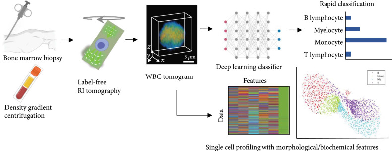
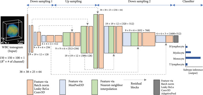
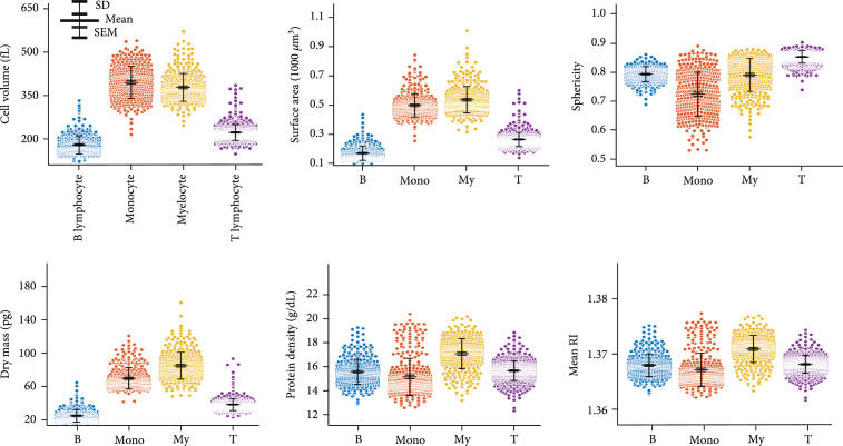
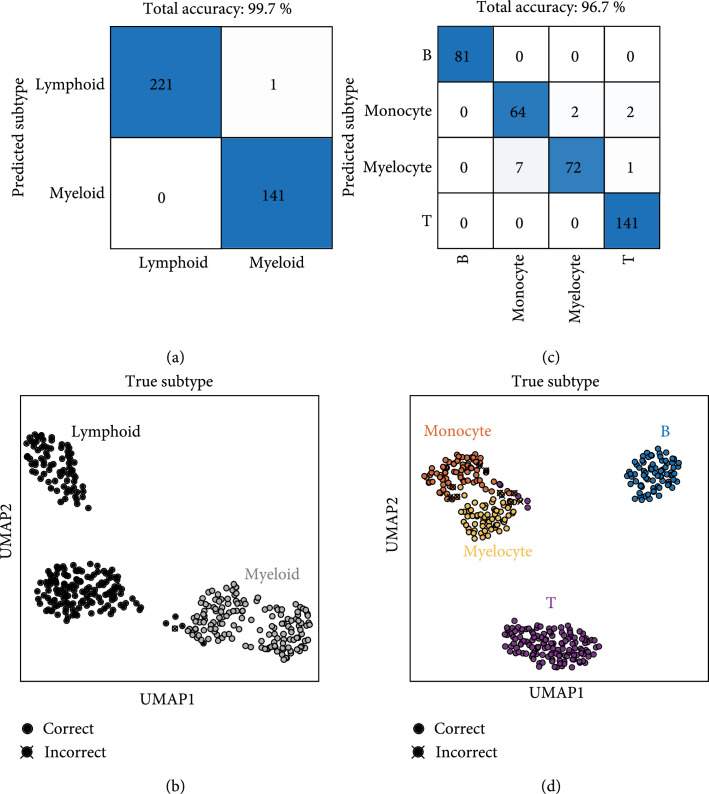
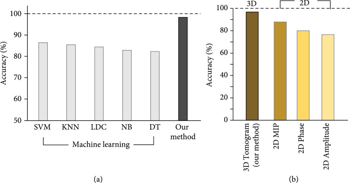
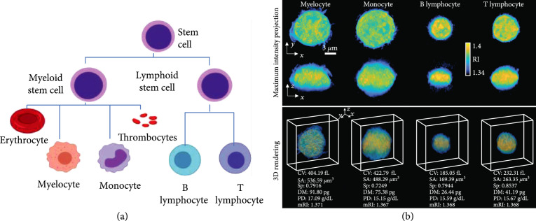
. We propose a rapid and accurate blood cell identification method exploiting deep learning and label-free refractive index (RI) tomography. Our computational approach that fully utilizes tomographic information of bone marrow (BM) white blood cell (WBC) enables us to not only classify the blood cells with deep learning but also quantitatively study their morphological and biochemical properties for hematology research. . Conventional methods for examining blood cells, such as blood smear analysis by medical professionals and fluorescence-activated cell sorting, require significant time, costs, and domain knowledge that could affect test results. While label-free imaging techniques that use a specimen's intrinsic contrast (e.g., multiphoton and Raman microscopy) have been used to characterize blood cells, their imaging procedures and instrumentations are relatively time-consuming and complex. . The RI tomograms of the BM WBCs are acquired via Mach-Zehnder interferometer-based tomographic microscope and classified by a 3D convolutional neural network. We test our deep learning classifier for the four types of bone marrow WBC collected from healthy donors (): monocyte, myelocyte, B lymphocyte, and T lymphocyte. The quantitative parameters of WBC are directly obtained from the tomograms. . Our results show >99% accuracy for the binary classification of myeloids and lymphoids and >96% accuracy for the four-type classification of B and T lymphocytes, monocyte, and myelocytes. The feature learning capability of our approach is visualized via an unsupervised dimension reduction technique. . We envision that the proposed cell classification framework can be easily integrated into existing blood cell investigation workflows, providing cost-effective and rapid diagnosis for hematologic malignancy.
我们提出了一种利用深度学习和无标记折射率(RI)断层扫描技术的快速准确的血细胞识别方法。我们的计算方法充分利用了骨髓(BM)白细胞(WBC)的断层扫描信息,不仅能够通过深度学习对血细胞进行分类,还能对其形态和生化特性进行定量研究,以用于血液学研究。传统的血细胞检测方法,如医学专业人员进行的血涂片分析和荧光激活细胞分选,需要大量时间、成本以及可能影响检测结果的专业知识。虽然使用样本固有对比度的无标记成像技术(如多光子和拉曼显微镜)已被用于表征血细胞,但其成像过程和仪器相对耗时且复杂。BM白细胞的RI断层图像通过基于马赫 - 曾德尔干涉仪的断层显微镜获取,并由三维卷积神经网络进行分类。我们对从健康供体收集的四种类型的骨髓白细胞(单核细胞、髓细胞、B淋巴细胞和T淋巴细胞)测试了我们的深度学习分类器。白细胞的定量参数直接从断层图像中获得。我们的结果表明,髓系和淋巴系的二元分类准确率>99%,B和T淋巴细胞、单核细胞和髓细胞的四元分类准确率>96%。我们通过无监督降维技术可视化了我们方法的特征学习能力。我们设想所提出的细胞分类框架可以轻松集成到现有的血细胞研究工作流程中,为血液系统恶性肿瘤提供经济高效且快速的诊断。