Service de Médecine et de Réadaptation Gériatrique et Neurologique, Hôpitaux de Saint-Maurice Saint-Maurice, France ; Inserm, U 1127, ICM FrontLab Paris, France ; CNRS, UMR 7225, ICM FrontLab Paris, France ; Sorbonne Universités, UPMC Univ Paris 06, UMRS 1127 Paris, France ; Institut du Cerveau et de la Moelle Épinière, ICM FrontLab Paris, France.
The Neuropsychological Laboratory, CNS-Fed Paris, France ; Laboratoire Psychologie de la Perception, UMR 8242 CNRS-Université Paris Descartes Paris, France.
Front Integr Neurosci. 2014 Sep 30;8:74. doi: 10.3389/fnint.2014.00074. eCollection 2014.
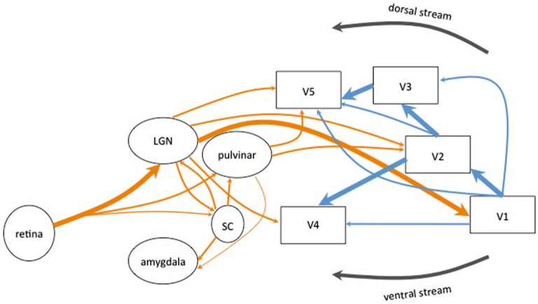
Visual field defects (VFDs) are one of the most common consequences observed after brain injury, especially after a stroke in the posterior cerebral artery territory. Less frequently, tumors, traumatic brain injury, brain surgery or demyelination can also determine various visual disabilities, from a decrease in visual acuity to cerebral blindness. Visual field defects is a factor of bad functional prognosis as it compromises many daily life activities (e.g., obstacle avoidance, driving, and reading) and therefore the patient's quality of life. Spontaneous recovery seems to be limited and restricted to the first 6 months, with the best chance of improvement at 1 month. The possible mechanisms at work could be partly due to cortical reorganization in the visual areas (plasticity) and/or partly to the use of intact alternative visual routes, first identified in animal studies and possibly underlying the phenomenon of blindsight. Despite processes of early recovery, which is rarely complete, and learning of compensatory strategies, the patient's autonomy may still be compromised at more chronic stages. Therefore, various rehabilitation therapies based on neuroanatomical knowledge have been developed to improve VFDs. These use eye-movement training techniques (e.g., visual search, saccadic eye movements), reading training, visual field restitution (the Vision Restoration Therapy, VRT), or perceptual learning. In this review, we will focus on studies of human adults with acquired VFDs, which have used different imaging techniques (Positron Emission Tomography, PET; Diffusion Tensor Imaging, DTI; functional Magnetic Resonance Imaging, fMRI; Magneto Encephalography, MEG) or neurostimulation techniques (Transcranial Magnetic Stimulation, TMS; transcranial Direct Current Stimulation, tDCS) to show brain activations in the course of spontaneous recovery or after specific rehabilitation techniques.
视野缺损(VFD)是脑损伤后最常见的后果之一,尤其是在后脑动脉区域发生中风后。较少见的是,肿瘤、创伤性脑损伤、脑外科手术或脱髓鞘也可能导致各种视力障碍,从视力下降到脑盲不等。视野缺损是功能预后不良的一个因素,因为它会影响许多日常生活活动(例如,避免障碍物、驾驶和阅读),从而影响患者的生活质量。自发恢复似乎是有限的,并且仅限于前 6 个月,在 1 个月时改善的机会最大。可能的工作机制部分归因于视觉区域的皮质重组(可塑性),或部分归因于对完整替代视觉途径的利用,首先在动物研究中发现,可能是盲视现象的基础。尽管存在早期恢复过程(很少完全恢复)和补偿策略的学习,但在更慢性阶段,患者的自主性仍可能受到影响。因此,基于神经解剖学知识的各种康复治疗方法已经被开发出来,以改善 VFD。这些方法使用眼球运动训练技术(例如,视觉搜索、眼跳运动)、阅读训练、视野恢复(视觉恢复疗法,VRT)或知觉学习。在这篇综述中,我们将重点关注使用不同成像技术(正电子发射断层扫描,PET;弥散张量成像,DTI;功能磁共振成像,fMRI;磁脑电图,MEG)或神经刺激技术(经颅磁刺激,TMS;经颅直流电刺激,tDCS)的成人后天 VFD 研究,以显示自发恢复过程中或特定康复技术后的大脑激活。