Lindner Diana, Li Jia, Savvatis Konstantinos, Klingel Karin, Blankenberg Stefan, Tschöpe Carsten, Westermann Dirk
Clinic for General and Interventional Cardiology, University Heart Center Hamburg, Martinistraße 52, 20246 Hamburg, Germany ; German Center for Cardiovascular Research (DZHK), Partner Sites, Hamburg/Kiel/Lübeck, Germany.
Clinic for General and Interventional Cardiology, University Heart Center Hamburg, Martinistraße 52, 20246 Hamburg, Germany.
Mediators Inflamm. 2014;2014:519528. doi: 10.1155/2014/519528. Epub 2014 Oct 13.
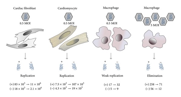
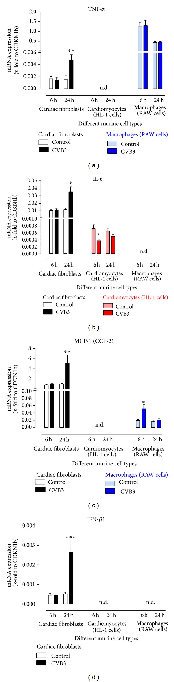
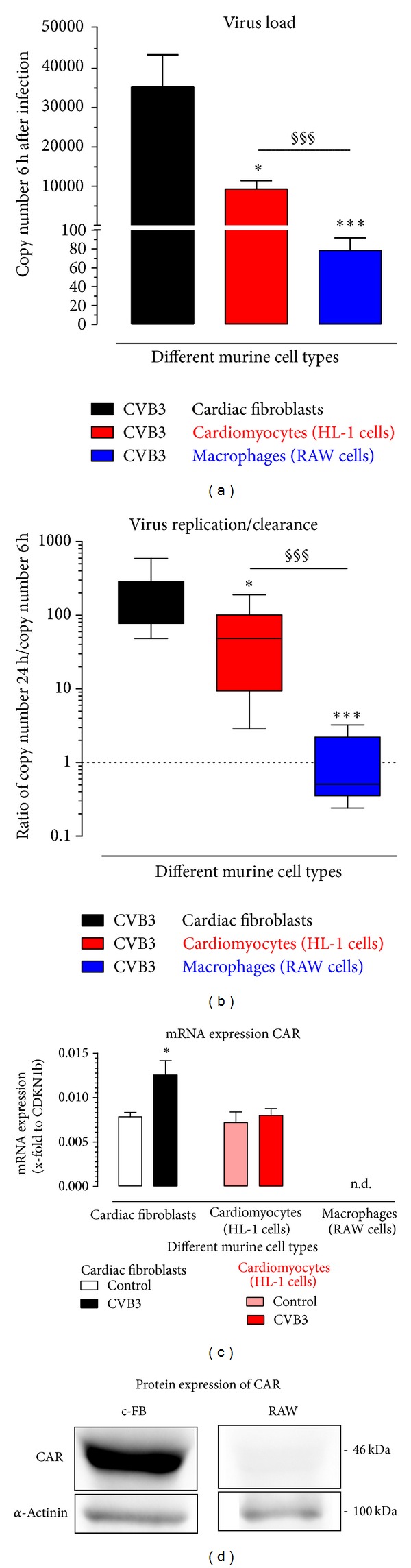
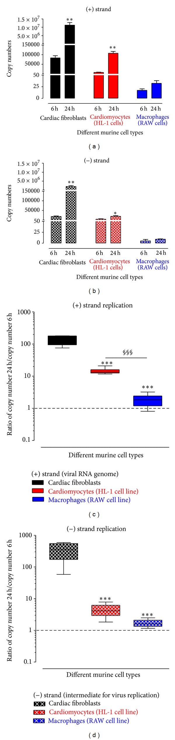
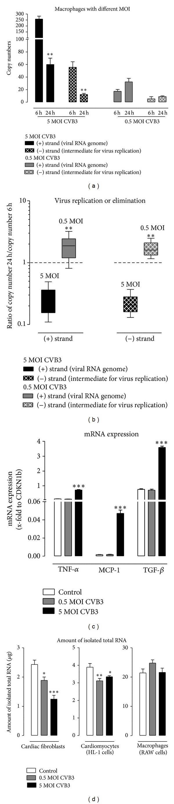
Myocarditis is an inflammatory disease caused by viral infection. Different subpopulations of leukocytes enter the cardiac tissue and lead to severe cardiac inflammation associated with myocyte loss and remodeling. Here, we study possible cell sources for viral replication using three compartments of the heart: fibroblasts, cardiomyocytes, and macrophages. We infected C57BL/6j mice with Coxsackievirus B3 (CVB3) and detected increased gene expression of anti-inflammatory and antiviral cytokines in the heart. Subsequently, we infected cardiac fibroblasts, cardiomyocytes, and macrophages with CVB3. Due to viral infection, the expression of TNF-α, IL-6, MCP-1, and IFN-β was significantly increased in cardiac fibroblasts compared to cardiomyocytes or macrophages. We found that in addition to cardiomyocytes cardiac fibroblasts were infected by CVB3 and displayed a higher virus replication (132-fold increase) compared to cardiomyocytes (14-fold increase) between 6 and 24 hours after infection. At higher virus concentrations, macrophages are able to reduce the viral copy number. At low virus concentration a persistent virus infection was determined. Therefore, we suggest that cardiac fibroblasts play an important role in the pathology of CVB3-induced myocarditis and are another important contributor of virus replication aggravating myocarditis.
心肌炎是一种由病毒感染引起的炎症性疾病。不同亚群的白细胞进入心脏组织,导致与心肌细胞丢失和重塑相关的严重心脏炎症。在此,我们利用心脏的三个组成部分:成纤维细胞、心肌细胞和巨噬细胞,研究病毒复制的可能细胞来源。我们用柯萨奇病毒B3(CVB3)感染C57BL/6j小鼠,并检测到心脏中抗炎和抗病毒细胞因子的基因表达增加。随后,我们用CVB3感染心脏成纤维细胞、心肌细胞和巨噬细胞。由于病毒感染,与心肌细胞或巨噬细胞相比,心脏成纤维细胞中TNF-α、IL-6、MCP-1和IFN-β的表达显著增加。我们发现,除心肌细胞外,心脏成纤维细胞也被CVB3感染,并且在感染后6至24小时之间,与心肌细胞(增加14倍)相比,显示出更高的病毒复制(增加132倍)。在较高病毒浓度下,巨噬细胞能够减少病毒拷贝数。在低病毒浓度下,确定存在持续性病毒感染。因此,我们认为心脏成纤维细胞在CVB3诱导的心肌炎病理过程中起重要作用,并且是加重心肌炎的病毒复制的另一个重要因素。