Tokunaga Kazuaki, Saitoh Noriko, Goldberg Ilya G, Sakamoto Chiyomi, Yasuda Yoko, Yoshida Yoshinori, Yamanaka Shinya, Nakao Mitsuyoshi
1] Department of Medical Cell Biology, Institute of Molecular Embryology and Genetics, Kumamoto University, 2-2-1 Honjo, Chuo-ku, Kumamoto 860-0811, Japan [2] Core Research for Evolutional Science and Technology (CREST), Japan Science and Technology Agency, Tokyo, Japan.
Image Informatics and Computational Biology Unit, Laboratory of Genetics, National Institute on Aging, National Institutes of Health, 251 Bayview Boulevard, Suite 100, Baltimore, MD 21224, USA.
Sci Rep. 2014 Nov 11;4:6996. doi: 10.1038/srep06996.
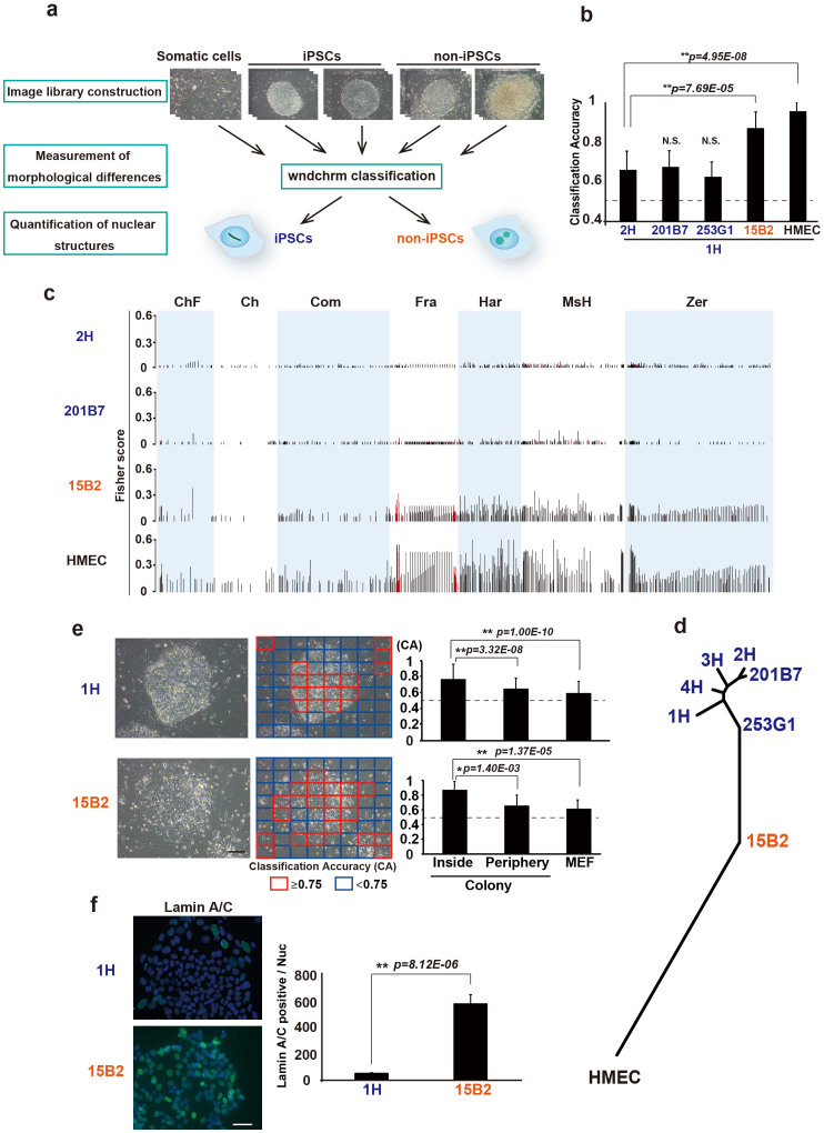
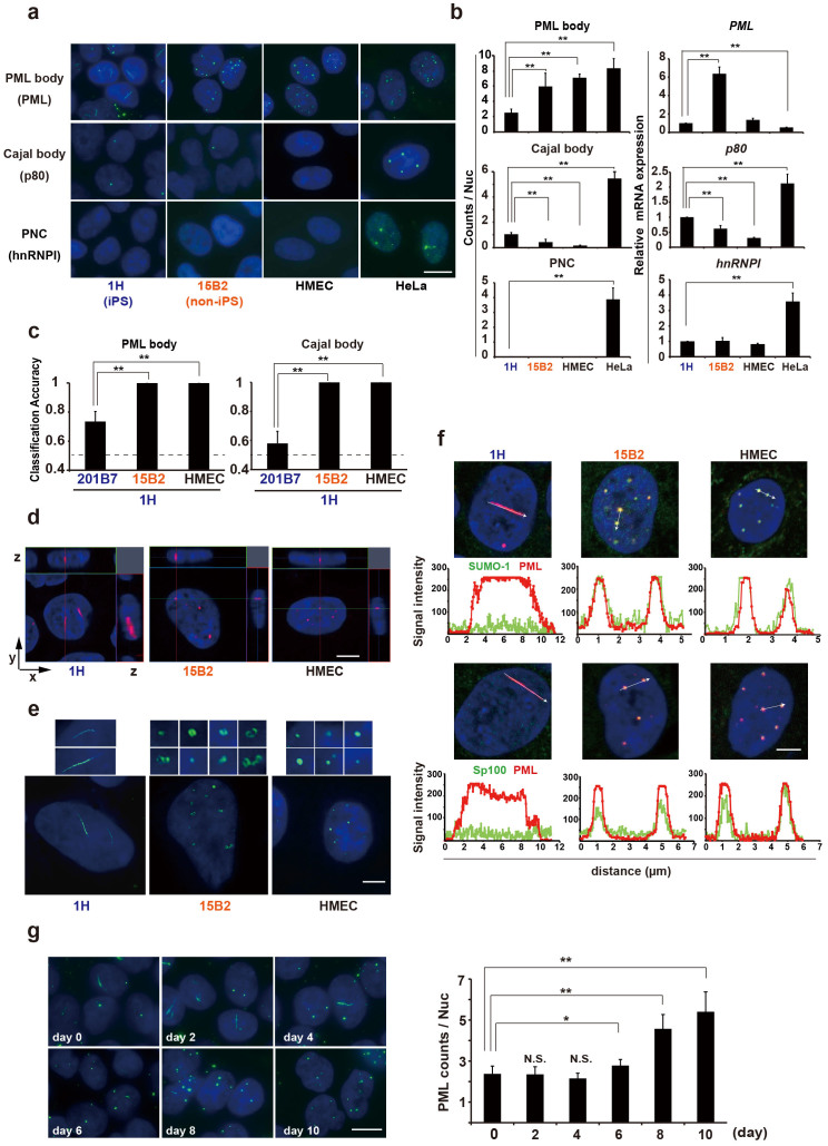
Non-invasive evaluation of cell reprogramming by advanced image analysis is required to maintain the quality of cells intended for regenerative medicine. Here, we constructed living and unlabelled colony image libraries of various human induced pluripotent stem cell (iPSC) lines for supervised machine learning pattern recognition to accurately distinguish bona fide iPSCs from improperly reprogrammed cells. Furthermore, we found that image features for efficient discrimination reside in cellular components. In fact, extensive analysis of nuclear morphologies revealed dynamic and characteristic signatures, including the linear form of the promyelocytic leukaemia (PML)-defined structure in iPSCs, which was reversed to a regular sphere upon differentiation. Our data revealed that iPSCs have a markedly different overall nuclear architecture that may contribute to highly accurate discrimination based on the cell reprogramming status.
为了维持用于再生医学的细胞质量,需要通过先进的图像分析对细胞重编程进行非侵入性评估。在此,我们构建了各种人类诱导多能干细胞(iPSC)系的活细胞和未标记的集落图像库,用于监督机器学习模式识别,以准确区分真正的iPSC与重编程不当的细胞。此外,我们发现有效区分的图像特征存在于细胞成分中。事实上,对核形态的广泛分析揭示了动态和特征性特征,包括iPSC中早幼粒细胞白血病(PML)定义结构的线性形式,其在分化时转变为规则的球体。我们的数据表明,iPSC具有明显不同的整体核结构,这可能有助于基于细胞重编程状态进行高度准确的区分。