Adam Yoav, Livneh Yoav, Miyamichi Kazunari, Groysman Maya, Luo Liqun, Mizrahi Adi
Department of Neurobiology, Institute of Life Sciences, The Edmond and Lily Safra Center for Brain Sciences, The Hebrew University of Jerusalem Jerusalem, Israel.
Department of Biology, Howard Hughes Medical Institute, Stanford University Stanford, CA, USA.
Front Neural Circuits. 2014 Nov 4;8:129. doi: 10.3389/fncir.2014.00129. eCollection 2014.
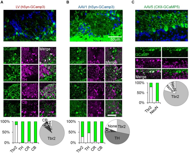
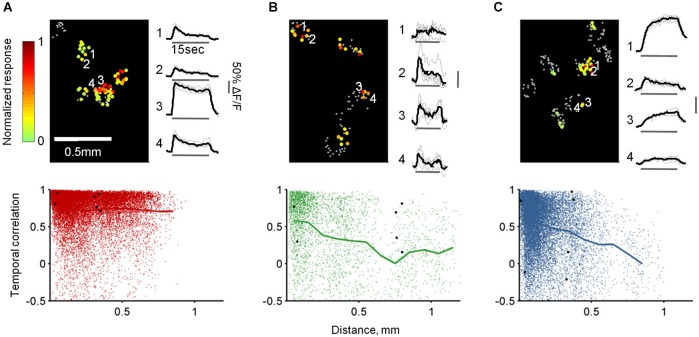
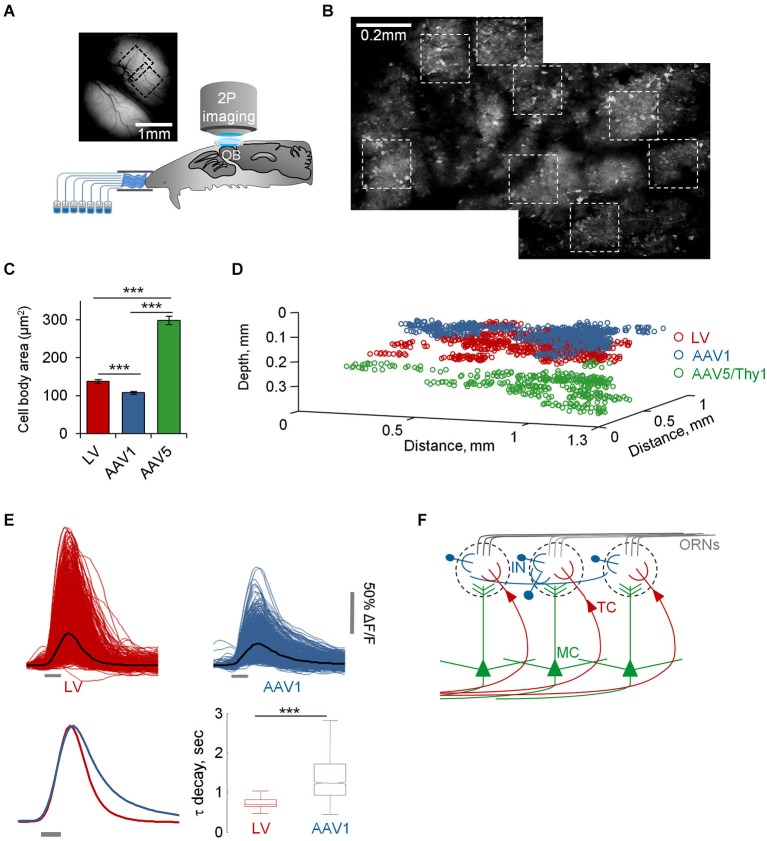
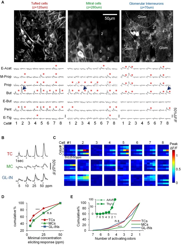
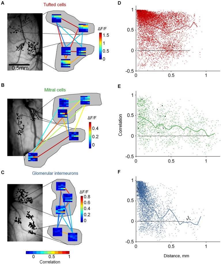
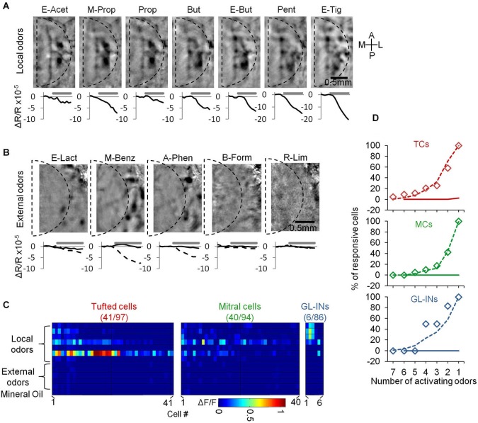
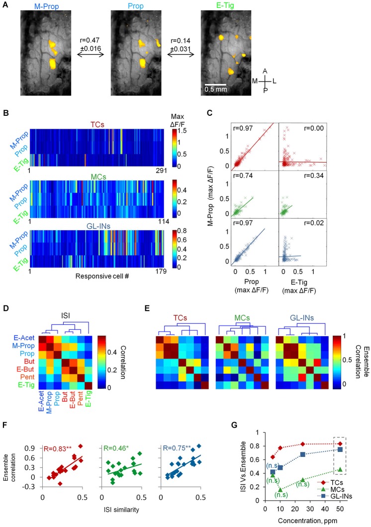
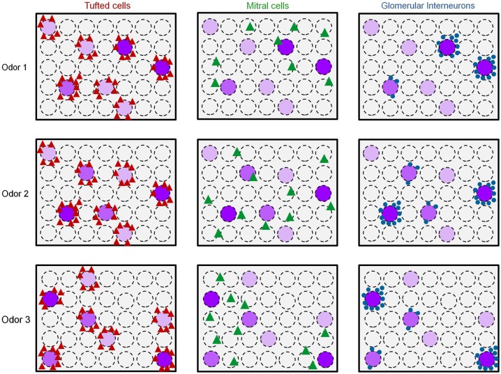
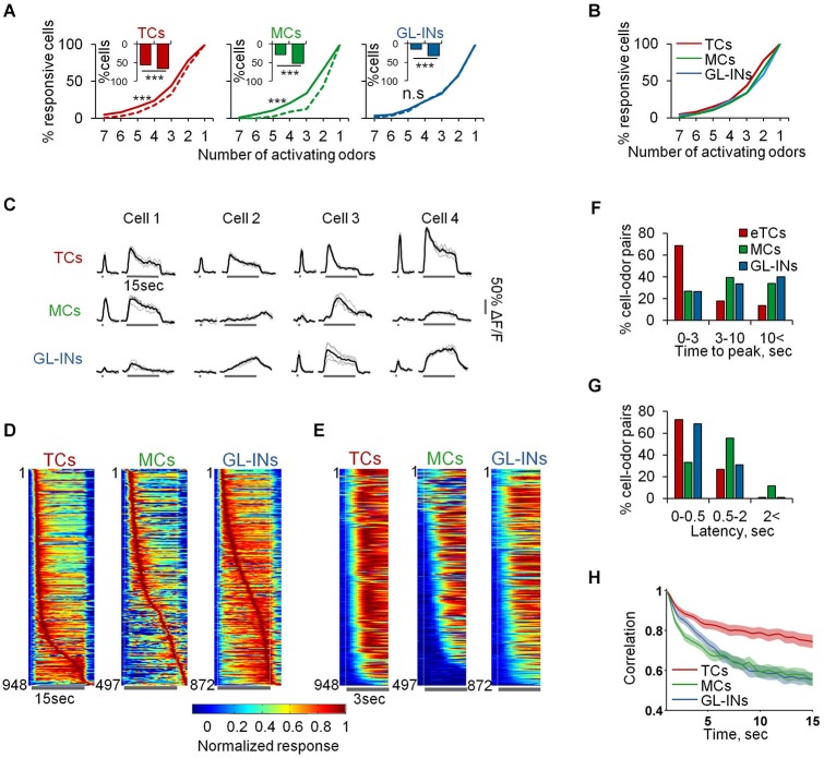
Sensory inputs from the nasal epithelium to the olfactory bulb (OB) are organized as a discrete map in the glomerular layer (GL). This map is then modulated by distinct types of local neurons and transmitted to higher brain areas via mitral and tufted cells. Little is known about the functional organization of the circuits downstream of glomeruli. We used in vivo two-photon calcium imaging for large scale functional mapping of distinct neuronal populations in the mouse OB, at single cell resolution. Specifically, we imaged odor responses of mitral cells (MCs), tufted cells (TCs) and glomerular interneurons (GL-INs). Mitral cells population activity was heterogeneous and only mildly correlated with the olfactory receptor neuron (ORN) inputs, supporting the view that discrete input maps undergo significant transformations at the output level of the OB. In contrast, population activity profiles of TCs were dense, and highly correlated with the odor inputs in both space and time. Glomerular interneurons were also highly correlated with the ORN inputs, but showed higher activation thresholds suggesting that these neurons are driven by strongly activated glomeruli. Temporally, upon persistent odor exposure, TCs quickly adapted. In contrast, both MCs and GL-INs showed diverse temporal response patterns, suggesting that GL-INs could contribute to the transformations MCs undergo at slow time scales. Our data suggest that sensory odor maps are transformed by TCs and MCs in different ways forming two distinct and parallel information streams.
从鼻上皮到嗅球(OB)的感觉输入在肾小球层(GL)中被组织成一个离散图谱。然后,这个图谱由不同类型的局部神经元进行调节,并通过二尖瓣细胞和簇状细胞传递到更高的脑区。关于肾小球下游回路的功能组织,我们所知甚少。我们使用体内双光子钙成像技术,以单细胞分辨率对小鼠嗅球中不同神经元群体进行大规模功能图谱绘制。具体而言,我们对二尖瓣细胞(MCs)、簇状细胞(TCs)和肾小球中间神经元(GL-INs)的气味反应进行了成像。二尖瓣细胞群体活动具有异质性,仅与嗅觉受体神经元(ORN)输入存在轻度相关性,这支持了离散输入图谱在嗅球输出水平会发生显著转变的观点。相比之下,簇状细胞的群体活动图谱密集,并且在空间和时间上都与气味输入高度相关。肾小球中间神经元也与ORN输入高度相关,但显示出更高的激活阈值,表明这些神经元由强烈激活的肾小球驱动。在时间上,持续暴露于气味时,簇状细胞会迅速适应。相比之下,二尖瓣细胞和肾小球中间神经元都表现出多样的时间反应模式,这表明肾小球中间神经元可能在缓慢的时间尺度上对二尖瓣细胞所经历的转变有所贡献。我们的数据表明,感觉气味图谱以不同方式由簇状细胞和二尖瓣细胞进行转变,形成两个不同且平行的信息流。