Department of Neuro- and Sensory Physiology, University of Göttingen Medical Center, European Neuroscience Institute, Cluster of Excellence Nanoscale Microscopy and Molecular Physiology of the Brain Göttingen, Germany ; International Max Planck Research School for Molecular Biology Göttingen, Germany.
Department of Neuro- and Sensory Physiology, University of Göttingen Medical Center, European Neuroscience Institute, Cluster of Excellence Nanoscale Microscopy and Molecular Physiology of the Brain Göttingen, Germany.
Front Cell Neurosci. 2014 Dec 8;8:409. doi: 10.3389/fncel.2014.00409. eCollection 2014.
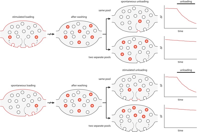
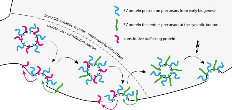
The trigger for synaptic vesicle exocytosis is Ca(2+), which enters the synaptic bouton following action potential stimulation. However, spontaneous release of neurotransmitter also occurs in the absence of stimulation in virtually all synaptic boutons. It has long been thought that this represents exocytosis driven by fluctuations in local Ca(2+) levels. The vesicles responding to these fluctuations are thought to be the same ones that release upon stimulation, albeit potentially triggered by different Ca(2+) sensors. This view has been challenged by several recent works, which have suggested that spontaneous release is driven by a separate pool of synaptic vesicles. Numerous articles appeared during the last few years in support of each of these hypotheses, and it has been challenging to bring them into accord. We speculate here on the origins of this controversy, and propose a solution that is related to developmental effects. Constitutive membrane traffic, needed for the biogenesis of vesicles and synapses, is responsible for high levels of spontaneous membrane fusion in young neurons, probably independent of Ca(2+). The vesicles releasing spontaneously in such neurons are not related to other synaptic vesicle pools and may represent constitutively releasing vesicles (CRVs) rather than bona fide synaptic vesicles. In mature neurons, constitutive traffic is much dampened, and the few remaining spontaneous release events probably represent bona fide spontaneously releasing synaptic vesicles (SRSVs) responding to Ca(2+) fluctuations, along with a handful of CRVs that participate in synaptic vesicle turnover.
引发突触小泡胞吐的是 Ca(2+),在动作电位刺激后,Ca(2+)进入突触末梢。然而,在几乎所有的突触末梢中,即使没有刺激也会发生神经递质的自发释放。长期以来,人们一直认为这代表了由局部 Ca(2+)水平波动驱动的胞吐作用。这些对波动作出反应的小泡被认为是在刺激时释放的小泡,尽管它们可能由不同的 Ca(2+)传感器触发。这一观点受到了最近几项研究的挑战,这些研究表明,自发释放是由一个单独的突触小泡池驱动的。在过去的几年里,出现了许多支持这些假说的文章,将它们协调一致是很有挑战性的。我们在这里推测了这一争议的起源,并提出了一个与发育效应有关的解决方案。对于小泡和突触的生物发生所需的组成型膜运输,是年轻神经元中自发膜融合的高水平的原因,可能与 Ca(2+)无关。在这些神经元中自发释放的小泡与其他突触小泡池无关,可能代表组成型释放小泡(CRVs),而不是真正的突触小泡。在成熟的神经元中,组成型运输受到了很大的抑制,而剩下的少数自发释放事件可能代表了真正的自发释放突触小泡(SRSVs)对 Ca(2+)波动的反应,以及少量参与突触小泡周转的 CRVs。