Ho Joseph, Adhi Mehreen, Baumal Caroline, Liu Jonathan, Fujimoto James G, Duker Jay S, Waheed Nadia K
*Department of Ophthalmology, New England Eye Center, Tufts Medical Center, Boston, Massachusetts; and †Department of Electrical Engineering and Computer Science, Research Laboratory of Electronics, Massachusetts Institute of Technology, Cambridge, Massachusetts.
Retina. 2015 Mar;35(3):467-72. doi: 10.1097/IAE.0000000000000355.
To assess the agreement and reproducibility of retinal pigment epithelial detachment (RPED) volumetric measurements using a commercially available optical coherence tomography software available for the Zeiss Cirrus HD-OCT.
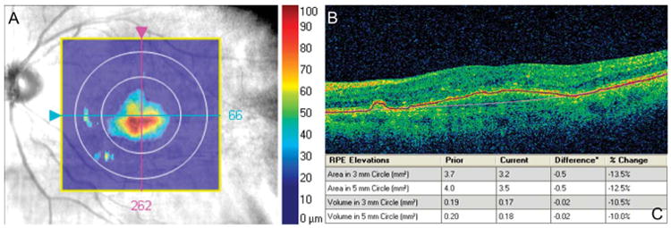
Twelve eyes of 10 patients with a diagnosis of neovascular age-related macular degeneration with RPED, seen at the New England Eye Center between October 2012 and December 2012, were enrolled in the study. Three separate scans per affected eye were obtained using the "Macular Cube 512 × 128" protocol. "Retinal pigment epithelial (RPE) elevation analysis" software was used to measure RPED volumes in the central 3-mm and 5-mm circles by calculating the volume between the "RPE fit" and "true RPE" lines. All 128 raster scans for each eye were exported into the AMIRA software for manual segmentation of RPED volumes in the central 3-mm and 5-mm circles. Interscan reproducibility and manual-to-automated agreement were assessed by intraclass correlation coefficient. Incidence of automated segmentation line error for both RPE fit and true RPE lines in the central 1 mm region was calculated.
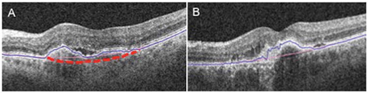
Average RPED volumes through automated segmentation software were 0.14 mm3 and 0.21 mm3 in the central 3-mm and 5-mm circles, respectively. Manual segmentation yielded average RPED volumes of 0.50 mm3 in the 3-mm circles and 0.92 mm3 in the 5-mm circles. Manual segmentation yielded significantly greater RPED volumes compared with automated measurements (P < 0.05). Intraclass correlation coefficients across the 3 automated measurements were 0.954 and 0.983 for volume in the 3-mm and 5-mm circles, respectively. Intraclass correlation coefficients between the manual and automatic volumes were 0.296 and 0.337 for the 3-mm and 5-mm circles, respectively. In the central 1 mm region, 11 of the 12 scans had breakdown in RPE fit line, whereas 8 of the 12 scans showed true RPE line breakdown.
Automated "RPED elevation" software demonstrated high interscan reproducibility. However, it showed low agreement with manual measurements from high rates of segmentation line breakdown, especially at the level of the RPE fit line (91.7%). Manual measurements resulted in greater volumes compared with automated measurements.
使用适用于蔡司Cirrus HD-OCT的市售光学相干断层扫描软件,评估视网膜色素上皮脱离(RPED)体积测量的一致性和可重复性。
2012年10月至2012年12月期间在新英格兰眼中心就诊的10例诊断为新生血管性年龄相关性黄斑变性伴RPED的患者的12只眼纳入研究。使用“黄斑立方体512×128”协议对每只患眼进行三次单独扫描。通过计算“RPE拟合”线和“真实RPE”线之间的体积,使用“视网膜色素上皮(RPE)隆起分析”软件测量中央3毫米和5毫米圆圈内的RPED体积。将每只眼的所有128条光栅扫描导出到AMIRA软件中,以手动分割中央3毫米和5毫米圆圈内的RPED体积。通过组内相关系数评估扫描间的可重复性和手动与自动测量结果的一致性。计算中央1毫米区域内RPE拟合线和真实RPE线的自动分割线误差发生率。
通过自动分割软件测得的中央3毫米和5毫米圆圈内RPED的平均体积分别为0.14立方毫米和0.21立方毫米。手动分割在3毫米圆圈内得到的RPED平均体积为0.50立方毫米,在5毫米圆圈内为0.92立方毫米。与自动测量相比,手动分割得到的RPED体积明显更大(P<0.05)。3次自动测量的组内相关系数在3毫米圆圈内的体积为0.954,在5毫米圆圈内为0.983。手动和自动测量体积之间的组内相关系数在3毫米圆圈内为0.296,在5毫米圆圈内为0.337。在中央1毫米区域,12次扫描中有11次RPE拟合线出现断裂,而12次扫描中有8次显示真实RPE线断裂。
自动“RPED隆起”软件显示出较高的扫描间可重复性。然而,由于分割线断裂率较高,尤其是在RPE拟合线水平(91.7%),它与手动测量的一致性较低。与自动测量相比,手动测量得到的体积更大。