Wang Ken, Lee Peter, Mirams Gary R, Sarathchandra Padmini, Borg Thomas K, Gavaghan David J, Kohl Peter, Bollensdorff Christian
Department of Computer Science, University of Oxford, Oxford, United Kingdom;
Department of Physics, University of Oxford, Clarendon Laboratory, Oxford, United Kingdom;
Am J Physiol Heart Circ Physiol. 2015 May 1;308(9):H1112-25. doi: 10.1152/ajpheart.00556.2014. Epub 2015 Jan 16.
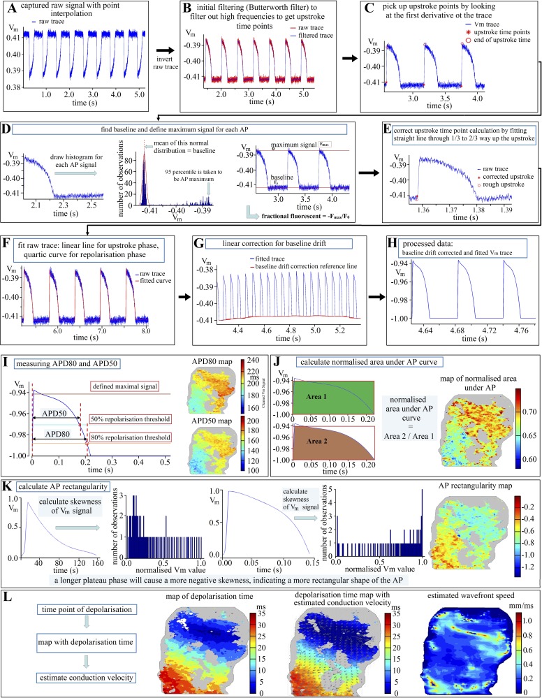
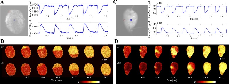
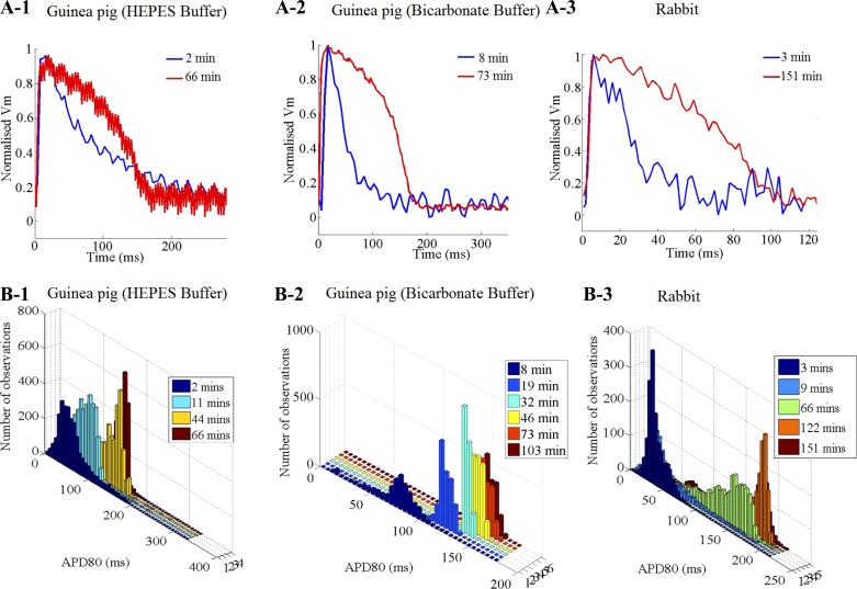
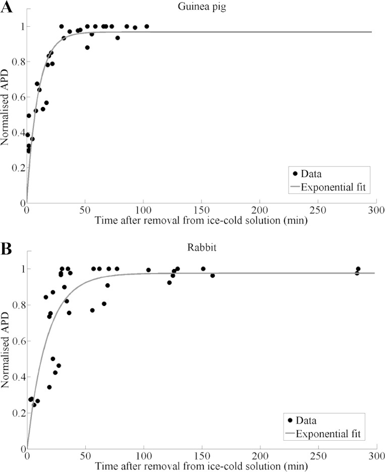
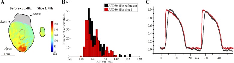
Cardiac tissue slices are becoming increasingly popular as a model system for cardiac electrophysiology and pharmacology research and development. Here, we describe in detail the preparation, handling, and optical mapping of transmembrane potential and intracellular free calcium concentration transients (CaT) in ventricular tissue slices from guinea pigs and rabbits. Slices cut in the epicardium-tangential plane contained well-aligned in-slice myocardial cell strands ("fibers") in subepicardial and midmyocardial sections. Cut with a high-precision slow-advancing microtome at a thickness of 350 to 400 μm, tissue slices preserved essential action potential (AP) properties of the precutting Langendorff-perfused heart. We identified the need for a postcutting recovery period of 36 min (guinea pig) and 63 min (rabbit) to reach 97.5% of final steady-state values for AP duration (APD) (identified by exponential fitting). There was no significant difference between the postcutting recovery dynamics in slices obtained using 2,3-butanedione 2-monoxime or blebistatin as electromechanical uncouplers during the cutting process. A rapid increase in APD, seen after cutting, was caused by exposure to ice-cold solution during the slicing procedure, not by tissue injury, differences in uncouplers, or pH-buffers (bicarbonate; HEPES). To characterize intrinsic patterns of CaT, AP, and conduction, a combination of multipoint and field stimulation should be used to avoid misinterpretation based on source-sink effects. In summary, we describe in detail the preparation, mapping, and data analysis approaches for reproducible cardiac tissue slice-based investigations into AP and CaT dynamics.
心脏组织切片作为心脏电生理学和药理学研究与开发的模型系统正变得越来越受欢迎。在此,我们详细描述了豚鼠和兔子心室组织切片跨膜电位和细胞内游离钙浓度瞬变(CaT)的制备、处理及光学映射。在心外膜切线平面切割的切片在下心外膜和心肌中层切片中包含排列良好的片内心肌细胞束(“纤维”)。用高精度慢速推进切片机以350至400μm的厚度切割,组织切片保留了切割前Langendorff灌注心脏的基本动作电位(AP)特性。我们确定需要36分钟(豚鼠)和63分钟(兔子)的切割后恢复期才能达到动作电位时程(APD)最终稳态值的97.5%(通过指数拟合确定)。在切割过程中使用2,3 - 丁二酮单肟或泡疹抑素作为机电解偶联剂获得的切片,其切割后恢复动力学之间没有显著差异。切割后观察到的APD快速增加是由切片过程中暴露于冰冷溶液引起的,而非组织损伤、解偶联剂差异或pH缓冲剂(碳酸氢盐;HEPES)所致。为了表征CaT、AP和传导的内在模式,应结合多点刺激和场刺激以避免基于源 - 汇效应的错误解读。总之,我们详细描述了基于心脏组织切片的可重复研究AP和CaT动力学的制备、映射及数据分析方法。