• 文献检索
  • 文档翻译
  • 深度研究
  • 学术资讯
  • Suppr Zotero 插件Zotero 插件
  • 邀请有礼
  • 套餐&价格
  • 历史记录
应用&插件
Suppr Zotero 插件Zotero 插件浏览器插件Mac 客户端Windows 客户端微信小程序
定价
高级版会员购买积分包购买API积分包
服务
文献检索文档翻译深度研究API 文档MCP 服务
关于我们
关于 Suppr公司介绍联系我们用户协议隐私条款
关注我们

Suppr 超能文献

核心技术专利:CN118964589B侵权必究
粤ICP备2023148730 号-1Suppr @ 2026

文献检索

告别复杂PubMed语法,用中文像聊天一样搜索,搜遍4000万医学文献。AI智能推荐,让科研检索更轻松。

立即免费搜索

文件翻译

保留排版,准确专业,支持PDF/Word/PPT等文件格式,支持 12+语言互译。

免费翻译文档

深度研究

AI帮你快速写综述,25分钟生成高质量综述,智能提取关键信息,辅助科研写作。

立即免费体验

基于学习的从胎儿脑部超声图像预测孕周

Learning-based prediction of gestational age from ultrasound images of the fetal brain.

作者信息

Namburete Ana I L, Stebbing Richard V, Kemp Bryn, Yaqub Mohammad, Papageorghiou Aris T, Alison Noble J

机构信息

Institute of Biomedical Engineering, Department of Engineering Science, University of Oxford, Oxford, United Kingdom.

Institute of Biomedical Engineering, Department of Engineering Science, University of Oxford, Oxford, United Kingdom.

出版信息

Med Image Anal. 2015 Apr;21(1):72-86. doi: 10.1016/j.media.2014.12.006. Epub 2015 Jan 3.

DOI:10.1016/j.media.2014.12.006
PMID:25624045
原文链接:https://pmc.ncbi.nlm.nih.gov/articles/PMC4339204/
Abstract

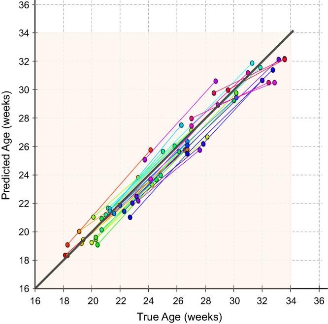
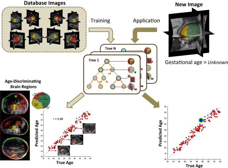
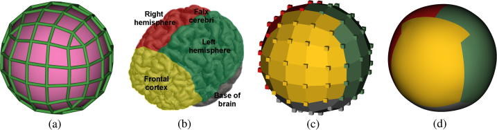
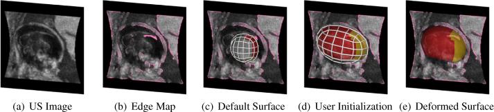
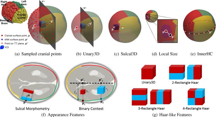
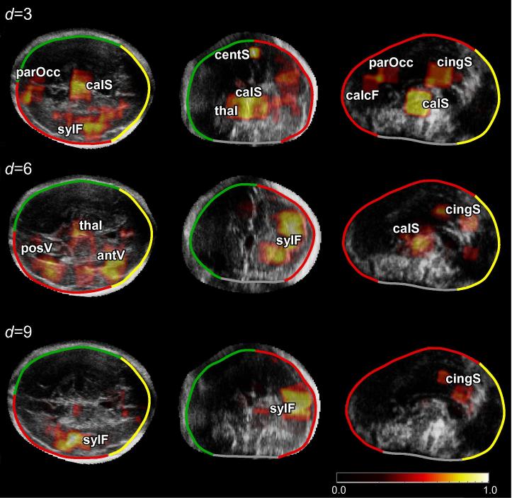
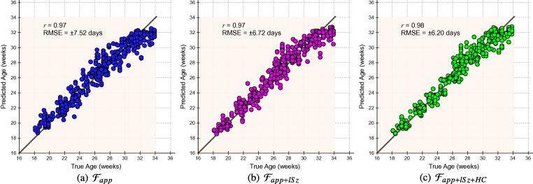
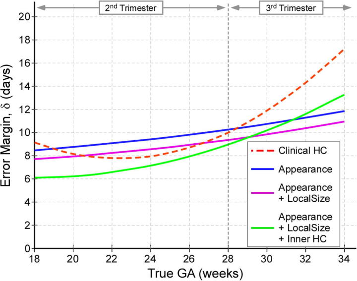
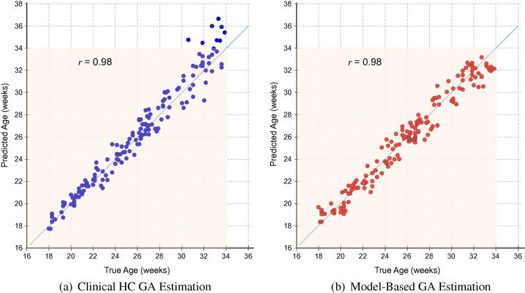
We propose an automated framework for predicting gestational age (GA) and neurodevelopmental maturation of a fetus based on 3D ultrasound (US) brain image appearance. Our method capitalizes on age-related sonographic image patterns in conjunction with clinical measurements to develop, for the first time, a predictive age model which improves on the GA-prediction potential of US images. The framework benefits from a manifold surface representation of the fetal head which delineates the inner skull boundary and serves as a common coordinate system based on cranial position. This allows for fast and efficient sampling of anatomically-corresponding brain regions to achieve like-for-like structural comparison of different developmental stages. We develop bespoke features which capture neurosonographic patterns in 3D images, and using a regression forest classifier, we characterize structural brain development both spatially and temporally to capture the natural variation existing in a healthy population (N=447) over an age range of active brain maturation (18-34weeks). On a routine clinical dataset (N=187) our age prediction results strongly correlate with true GA (r=0.98,accurate within±6.10days), confirming the link between maturational progression and neurosonographic activity observable across gestation. Our model also outperforms current clinical methods by ±4.57 days in the third trimester-a period complicated by biological variations in the fetal population. Through feature selection, the model successfully identified the most age-discriminating anatomies over this age range as being the Sylvian fissure, cingulate, and callosal sulci.

摘要

我们提出了一个基于三维超声(US)脑部图像外观预测胎儿孕周(GA)和神经发育成熟度的自动化框架。我们的方法利用与年龄相关的超声图像模式并结合临床测量,首次开发出一种预测年龄模型,该模型提高了超声图像预测孕周的潜力。该框架受益于胎儿头部的多流形表面表示,它描绘了内颅骨边界,并作为基于颅骨位置的通用坐标系。这使得能够快速有效地对解剖学上对应的脑区进行采样,以实现不同发育阶段的同类结构比较。我们开发了定制特征来捕捉三维图像中的神经超声模式,并使用回归森林分类器,在空间和时间上对脑结构发育进行表征,以捕捉健康人群(N = 447)在脑活跃成熟年龄范围(18 - 34周)内存在的自然变异。在一个常规临床数据集(N = 187)上,我们的年龄预测结果与实际孕周高度相关(r = 0.98,在±6.10天内准确),证实了整个孕期可观察到的成熟进展与神经超声活动之间的联系。在孕晚期(这一时期受胎儿群体生物学变异的影响),我们的模型在预测准确性上比当前临床方法提高了±4.57天。通过特征选择,该模型成功识别出在这个年龄范围内最能区分年龄的解剖结构为外侧裂、扣带回和胼胝体沟。

https://cdn.ncbi.nlm.nih.gov/pmc/blobs/9b89/4339204/46634f259a49/gr13.jpg
https://cdn.ncbi.nlm.nih.gov/pmc/blobs/9b89/4339204/a431438737b7/fx1.jpg
https://cdn.ncbi.nlm.nih.gov/pmc/blobs/9b89/4339204/416865bb2af8/gr1.jpg
https://cdn.ncbi.nlm.nih.gov/pmc/blobs/9b89/4339204/cf234cc8a4fc/gr2.jpg
https://cdn.ncbi.nlm.nih.gov/pmc/blobs/9b89/4339204/d01061cf82bc/gr3.jpg
https://cdn.ncbi.nlm.nih.gov/pmc/blobs/9b89/4339204/6e9eac3156dc/gr4.jpg
https://cdn.ncbi.nlm.nih.gov/pmc/blobs/9b89/4339204/395f19d263a3/gr5.jpg
https://cdn.ncbi.nlm.nih.gov/pmc/blobs/9b89/4339204/60ec96e83aa6/gr6.jpg
https://cdn.ncbi.nlm.nih.gov/pmc/blobs/9b89/4339204/7664456a9925/gr7.jpg
https://cdn.ncbi.nlm.nih.gov/pmc/blobs/9b89/4339204/362c35fd8f0c/gr8.jpg
https://cdn.ncbi.nlm.nih.gov/pmc/blobs/9b89/4339204/88c82f602cdc/gr9.jpg
https://cdn.ncbi.nlm.nih.gov/pmc/blobs/9b89/4339204/6950ebecbae5/gr10.jpg
https://cdn.ncbi.nlm.nih.gov/pmc/blobs/9b89/4339204/cc12469ca13f/gr11.jpg
https://cdn.ncbi.nlm.nih.gov/pmc/blobs/9b89/4339204/63efef6bafb8/gr12.jpg
https://cdn.ncbi.nlm.nih.gov/pmc/blobs/9b89/4339204/46634f259a49/gr13.jpg

相似文献

1
Learning-based prediction of gestational age from ultrasound images of the fetal brain.基于学习的从胎儿脑部超声图像预测孕周
Med Image Anal. 2015 Apr;21(1):72-86. doi: 10.1016/j.media.2014.12.006. Epub 2015 Jan 3.
2
Predicting fetal neurodevelopmental age from ultrasound images.从超声图像预测胎儿神经发育年龄。
Med Image Comput Comput Assist Interv. 2014;17(Pt 2):260-7. doi: 10.1007/978-3-319-10470-6_33.
3
Detection and measurement of fetal anatomies from ultrasound images using a constrained probabilistic boosting tree.使用约束概率提升树从超声图像中检测和测量胎儿解剖结构。
IEEE Trans Med Imaging. 2008 Sep;27(9):1342-55. doi: 10.1109/TMI.2008.928917.
4
Registration of 3D fetal neurosonography and MRI.三维胎儿神经超声和 MRI 注册。
Med Image Anal. 2013 Dec;17(8):1137-50. doi: 10.1016/j.media.2013.07.004. Epub 2013 Jul 30.
5
Analysis of maturation features in fetal brain ultrasound via artificial intelligence for the estimation of gestational age.人工智能分析胎儿大脑超声的成熟特征,用于估计胎龄。
Am J Obstet Gynecol MFM. 2021 Nov;3(6):100462. doi: 10.1016/j.ajogmf.2021.100462. Epub 2021 Aug 14.
6
Automatic segmentation of the cerebellum of fetuses on 3D ultrasound images, using a 3D Point Distribution Model.使用三维点分布模型对三维超声图像上的胎儿小脑进行自动分割。
Annu Int Conf IEEE Eng Med Biol Soc. 2010;2010:4731-4. doi: 10.1109/IEMBS.2010.5626624.
7
A fast automatic recognition and location algorithm for fetal genital organs in ultrasound images.一种用于超声图像中胎儿生殖器官的快速自动识别与定位算法。
J Zhejiang Univ Sci B. 2009 Sep;10(9):648-58. doi: 10.1631/jzus.B0930162.
8
Registration of 3D fetal brain US and MRI.三维胎儿脑部超声和磁共振成像的配准
Med Image Comput Comput Assist Interv. 2012;15(Pt 2):667-74. doi: 10.1007/978-3-642-33418-4_82.
9
Ultrasound-based gestational-age estimation in late pregnancy.孕晚期基于超声的孕周估计
Ultrasound Obstet Gynecol. 2016 Dec;48(6):719-726. doi: 10.1002/uog.15894.
10
Automatic staging of placental maturity based on dense descriptor.基于密集描述符的胎盘成熟度自动分期。
Biomed Mater Eng. 2014;24(6):2821-9. doi: 10.3233/BME-141100.

引用本文的文献

1
Use of artificial intelligence for gestational age estimation: a systematic review and meta-analysis.人工智能在估计孕周中的应用:一项系统评价和荟萃分析。
Front Glob Womens Health. 2025 Jan 30;6:1447579. doi: 10.3389/fgwh.2025.1447579. eCollection 2025.
2
Repeatability and reproducibility of artificial intelligence-acquired fetal brain measurements (SonoCNS) in the second and third trimesters of pregnancy.人工智能获取胎儿脑测量值(SonoCNS)在妊娠第二和第三 trimester 的可重复性和再现性。
Sci Rep. 2024 Oct 23;14(1):25076. doi: 10.1038/s41598-024-77313-w.
3
Advancements in Artificial Intelligence for Fetal Neurosonography: A Comprehensive Review.

本文引用的文献

1
Predicting fetal neurodevelopmental age from ultrasound images.从超声图像预测胎儿神经发育年龄。
Med Image Comput Comput Assist Interv. 2014;17(Pt 2):260-7. doi: 10.1007/978-3-319-10470-6_33.
2
International standards for fetal growth based on serial ultrasound measurements: the Fetal Growth Longitudinal Study of the INTERGROWTH-21st Project.基于系列超声测量的胎儿生长国际标准:INTERGROWTH-21st 项目胎儿生长纵向研究。
Lancet. 2014 Sep 6;384(9946):869-79. doi: 10.1016/S0140-6736(14)61490-2.
3
Automatic quantification of normal cortical folding patterns from fetal brain MRI.
胎儿神经超声检查中人工智能的进展:综述
J Clin Med. 2024 Sep 22;13(18):5626. doi: 10.3390/jcm13185626.
4
A benchmark for 2D foetal brain ultrasound analysis.二维胎儿脑超声分析基准
Sci Data. 2024 Aug 24;11(1):923. doi: 10.1038/s41597-024-03774-3.
5
Trajectories of brain and behaviour development in the womb, at birth and through infancy.子宫内、出生时和婴儿期的大脑和行为发育轨迹。
Nat Hum Behav. 2024 Jul;8(7):1251-1262. doi: 10.1038/s41562-024-01896-7. Epub 2024 Jun 17.
6
The role of cortical structural variance in deep learning-based prediction of fetal brain age.皮质结构变异在基于深度学习的胎儿脑龄预测中的作用。
Front Neurosci. 2024 May 23;18:1411334. doi: 10.3389/fnins.2024.1411334. eCollection 2024.
7
A deep learning framework for noninvasive fetal ECG signal extraction.一种用于无创胎儿心电图信号提取的深度学习框架。
Front Physiol. 2024 Apr 22;15:1329313. doi: 10.3389/fphys.2024.1329313. eCollection 2024.
8
Evolving the Era of 5D Ultrasound? A Systematic Literature Review on the Applications for Artificial Intelligence Ultrasound Imaging in Obstetrics and Gynecology.开启5D超声时代?关于人工智能超声成像在妇产科应用的系统文献综述
J Clin Med. 2023 Oct 29;12(21):6833. doi: 10.3390/jcm12216833.
9
Normative spatiotemporal fetal brain maturation with satisfactory development at 2 years.正常胎儿脑成熟的时空模式,2 岁时发育良好。
Nature. 2023 Nov;623(7985):106-114. doi: 10.1038/s41586-023-06630-3. Epub 2023 Oct 25.
10
Application and Progress of Artificial Intelligence in Fetal Ultrasound.人工智能在胎儿超声中的应用与进展
J Clin Med. 2023 May 5;12(9):3298. doi: 10.3390/jcm12093298.
从胎儿脑磁共振成像中自动定量分析正常皮质折叠模式。
Neuroimage. 2014 May 1;91:21-32. doi: 10.1016/j.neuroimage.2014.01.034. Epub 2014 Jan 25.
4
A spatio-temporal latent atlas for semi-supervised learning of fetal brain segmentations and morphological age estimation.时空潜在图谱用于半监督学习胎儿脑分割和形态年龄估计。
Med Image Anal. 2014 Jan;18(1):9-21. doi: 10.1016/j.media.2013.08.004. Epub 2013 Aug 30.
5
Registration of 3D fetal neurosonography and MRI.三维胎儿神经超声和 MRI 注册。
Med Image Anal. 2013 Dec;17(8):1137-50. doi: 10.1016/j.media.2013.07.004. Epub 2013 Jul 30.
6
Neighbourhood approximation using randomized forests.基于随机森林的邻域逼近。
Med Image Anal. 2013 Oct;17(7):790-804. doi: 10.1016/j.media.2013.04.013. Epub 2013 May 10.
7
Development of the fetal cerebral cortex in the second trimester: assessment with 7T postmortem MR imaging.胎儿大脑皮质在妊娠中期的发育:7T 后尸体磁共振成像评估。
AJNR Am J Neuroradiol. 2013 Jul;34(7):1462-7. doi: 10.3174/ajnr.A3406. Epub 2013 Feb 14.
8
A feature-based developmental model of the infant brain in structural MRI.基于特征的婴儿大脑结构MRI发育模型。
Med Image Comput Comput Assist Interv. 2012;15(Pt 2):204-11. doi: 10.1007/978-3-642-33418-4_26.
9
The relevance voxel machine (RVoxM): a self-tuning Bayesian model for informative image-based prediction.相关性体素机(RVoxM):一种用于基于图像的信息预测的自调谐贝叶斯模型。
IEEE Trans Med Imaging. 2012 Dec;31(12):2290-306. doi: 10.1109/TMI.2012.2216543. Epub 2012 Sep 19.
10
Brain maturation: predicting individual BrainAGE in children and adolescents using structural MRI.脑成熟度:使用结构 MRI 预测儿童和青少年的个体脑龄。
Neuroimage. 2012 Nov 15;63(3):1305-12. doi: 10.1016/j.neuroimage.2012.08.001. Epub 2012 Aug 11.