• 文献检索
  • 文档翻译
  • 深度研究
  • 学术资讯
  • Suppr Zotero 插件Zotero 插件
  • 邀请有礼
  • 套餐&价格
  • 历史记录
应用&插件
Suppr Zotero 插件Zotero 插件浏览器插件Mac 客户端Windows 客户端微信小程序
定价
高级版会员购买积分包购买API积分包
服务
文献检索文档翻译深度研究API 文档MCP 服务
关于我们
关于 Suppr公司介绍联系我们用户协议隐私条款
关注我们

Suppr 超能文献

核心技术专利:CN118964589B侵权必究
粤ICP备2023148730 号-1Suppr @ 2026

文献检索

告别复杂PubMed语法,用中文像聊天一样搜索,搜遍4000万医学文献。AI智能推荐,让科研检索更轻松。

立即免费搜索

文件翻译

保留排版,准确专业,支持PDF/Word/PPT等文件格式,支持 12+语言互译。

免费翻译文档

深度研究

AI帮你快速写综述,25分钟生成高质量综述,智能提取关键信息,辅助科研写作。

立即免费体验

三维胎儿神经超声和 MRI 注册。

Registration of 3D fetal neurosonography and MRI.

机构信息

Institute of Biomedical Engineering, Department of Engineering Science, University of Oxford, UK; Department of Biomedical Engineering, King's College London, UK; Centre for the Developing Brain, King's College London, UK.

出版信息

Med Image Anal. 2013 Dec;17(8):1137-50. doi: 10.1016/j.media.2013.07.004. Epub 2013 Jul 30.

DOI:10.1016/j.media.2013.07.004
PMID:23969169
原文链接:https://pmc.ncbi.nlm.nih.gov/articles/PMC3807810/
Abstract

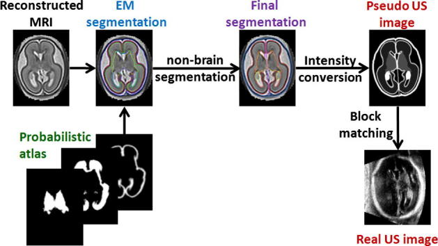
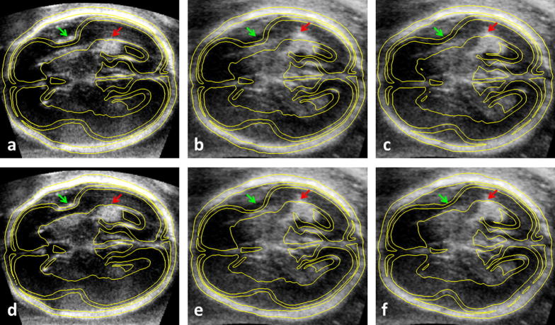
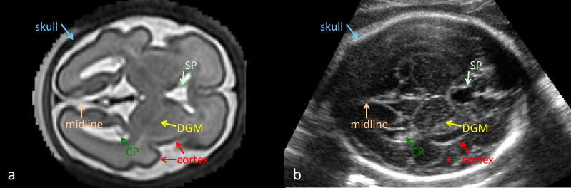
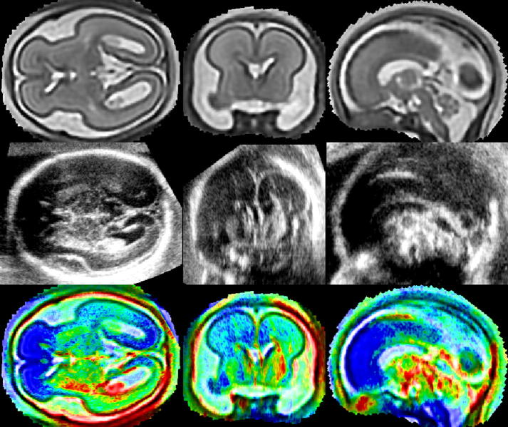
We propose a method for registration of 3D fetal brain ultrasound with a reconstructed magnetic resonance fetal brain volume. This method, for the first time, allows the alignment of models of the fetal brain built from magnetic resonance images with 3D fetal brain ultrasound, opening possibilities to develop new, prior information based image analysis methods for 3D fetal neurosonography. The reconstructed magnetic resonance volume is first segmented using a probabilistic atlas and a pseudo ultrasound image volume is simulated from the segmentation. This pseudo ultrasound image is then affinely aligned with clinical ultrasound fetal brain volumes using a robust block-matching approach that can deal with intensity artefacts and missing features in the ultrasound images. A qualitative and quantitative evaluation demonstrates good performance of the method for our application, in comparison with other tested approaches. The intensity average of 27 ultrasound images co-aligned with the pseudo ultrasound template shows good correlation with anatomy of the fetal brain as seen in the reconstructed magnetic resonance image.

摘要

我们提出了一种将 3D 胎儿脑超声与重建的磁共振胎儿脑容积配准的方法。该方法首次允许从磁共振图像构建的胎儿脑模型与 3D 胎儿脑超声进行配准,为开发新的基于先验信息的 3D 胎儿神经超声图像分析方法开辟了可能性。首先使用概率图谱对重建的磁共振容积进行分割,并从分割中模拟出伪超声图像容积。然后,使用稳健的块匹配方法将伪超声图像与临床超声胎儿脑容积进行仿射对齐,该方法可以处理超声图像中的强度伪影和缺失特征。定性和定量评估表明,与其他测试方法相比,该方法在我们的应用中具有良好的性能。与伪超声模板共配准的 27 张超声图像的强度平均值与重建磁共振图像中所见的胎儿脑解剖结构具有很好的相关性。

https://cdn.ncbi.nlm.nih.gov/pmc/blobs/388a/3807810/3896ba17e887/gr8.jpg
https://cdn.ncbi.nlm.nih.gov/pmc/blobs/388a/3807810/d88dc4b0dc8f/fx1.jpg
https://cdn.ncbi.nlm.nih.gov/pmc/blobs/388a/3807810/935369d4a9fe/gr2.jpg
https://cdn.ncbi.nlm.nih.gov/pmc/blobs/388a/3807810/60fbc9c45754/gr3.jpg
https://cdn.ncbi.nlm.nih.gov/pmc/blobs/388a/3807810/90954bfa3491/gr4.jpg
https://cdn.ncbi.nlm.nih.gov/pmc/blobs/388a/3807810/f69d434883c5/gr6.jpg
https://cdn.ncbi.nlm.nih.gov/pmc/blobs/388a/3807810/3896ba17e887/gr8.jpg

相似文献

1
Registration of 3D fetal neurosonography and MRI.三维胎儿神经超声和 MRI 注册。
Med Image Anal. 2013 Dec;17(8):1137-50. doi: 10.1016/j.media.2013.07.004. Epub 2013 Jul 30.
2
Registration of 3D fetal brain US and MRI.三维胎儿脑部超声和磁共振成像的配准
Med Image Comput Comput Assist Interv. 2012;15(Pt 2):667-74. doi: 10.1007/978-3-642-33418-4_82.
3
Automatic ultrasound-MRI registration for neurosurgery using the 2D and 3D LC(2) Metric.基于二维和三维 LC(2)度量的神经外科自动超声-MRI 配准。
Med Image Anal. 2014 Dec;18(8):1312-9. doi: 10.1016/j.media.2014.04.008. Epub 2014 May 2.
4
Intraoperative patient registration using volumetric true 3D ultrasound without fiducials.术中无基准点容积式真实 3D 超声患者注册。
Med Phys. 2012 Dec;39(12):7540-52. doi: 10.1118/1.4767758.
5
Segmentation of embryonic and fetal 3D ultrasound images based on pixel intensity distributions and shape priors.基于像素强度分布和形状先验的胚胎和胎儿 3D 超声图像分割。
Med Image Anal. 2015 Aug;24(1):255-268. doi: 10.1016/j.media.2014.12.005. Epub 2015 Jan 10.
6
Atlas-based segmentation of the germinal matrix from in utero clinical MRI of the fetal brain.基于图谱的胎儿脑宫内临床磁共振成像中生发基质的分割
Med Image Comput Comput Assist Interv. 2008;11(Pt 1):351-8. doi: 10.1007/978-3-540-85988-8_42.
7
Robust brain registration using adaptive probabilistic atlas.使用自适应概率图谱进行稳健的脑图谱配准。
Med Image Comput Comput Assist Interv. 2008;11(Pt 2):1041-9. doi: 10.1007/978-3-540-85990-1_125.
8
Non-rigid registration with missing correspondences in preoperative and postresection brain images.术前和切除术后脑图像中存在对应关系缺失的非刚性配准。
Med Image Comput Comput Assist Interv. 2010;13(Pt 1):367-74. doi: 10.1007/978-3-642-15705-9_45.
9
A unifying approach to registration, segmentation, and intensity correction.一种用于配准、分割和强度校正的统一方法。
Med Image Comput Comput Assist Interv. 2005;8(Pt 1):310-8. doi: 10.1007/11566465_39.
10
Characterization of carotid plaques on 3-dimensional ultrasound imaging by registration with multicontrast magnetic resonance imaging.基于多对比度磁共振成像配准的三维超声成像对颈动脉斑块的特征描述。
J Ultrasound Med. 2012 Oct;31(10):1567-80. doi: 10.7863/jum.2012.31.10.1567.

引用本文的文献

1
Towards automatic US-MR fetal brain image registration with learning-based methods.利用基于学习的方法实现超声-磁共振胎儿脑图像的自动配准
Neuroimage. 2025 Apr 15;310:121104. doi: 10.1016/j.neuroimage.2025.121104. Epub 2025 Mar 7.
2
A Systematic Review of Methodology Used in Studies Aimed at Creating Charts of Fetal Brain Structures.旨在创建胎儿脑结构图谱的研究中所使用方法的系统评价
Diagnostics (Basel). 2021 May 21;11(6):916. doi: 10.3390/diagnostics11060916.
3
Probabilistic Learning Coherent Point Drift for 3D Ultrasound Fetal Head Registration.

本文引用的文献

1
Registration of 3D fetal brain US and MRI.三维胎儿脑部超声和磁共振成像的配准
Med Image Comput Comput Assist Interv. 2012;15(Pt 2):667-74. doi: 10.1007/978-3-642-33418-4_82.
2
Reconstruction of fetal brain MRI with intensity matching and complete outlier removal.基于强度匹配和完全异常值去除的胎儿脑 MRI 重建。
Med Image Anal. 2012 Dec;16(8):1550-64. doi: 10.1016/j.media.2012.07.004. Epub 2012 Aug 9.
3
Prenatal diagnosis and outcome of fetal posterior fossa fluid collections.胎儿后颅窝液体积聚的产前诊断和结局。
基于概率学习的相干点漂移在三维超声胎儿头部配准中的应用。
Comput Math Methods Med. 2020 Jan 31;2020:4271519. doi: 10.1155/2020/4271519. eCollection 2020.
4
Analyzing the co-localization of substantia nigra hyper-echogenicities and iron accumulation in Parkinson's disease: A multi-modal atlas study with transcranial ultrasound and MRI.帕金森病中黑质高回声与铁沉积的共定位分析:一项经颅超声与磁共振成像的多模态图谱研究
Neuroimage Clin. 2020;26:102185. doi: 10.1016/j.nicl.2020.102185. Epub 2020 Feb 1.
5
Automatic and efficient MRI-US segmentations for improving intraoperative image fusion in image-guided neurosurgery.用于改善图像引导神经外科术中图像融合的自动高效 MRI-US 分割。
Neuroimage Clin. 2019;22:101766. doi: 10.1016/j.nicl.2019.101766. Epub 2019 Mar 12.
6
Fetal neuroimaging: an update on technical advances and clinical findings.胎儿神经影像学:技术进展与临床发现的最新情况
Pediatr Radiol. 2018 Apr;48(4):471-485. doi: 10.1007/s00247-017-3965-z. Epub 2018 Mar 17.
7
Nonlinear deformation of tractography in ultrasound-guided low-grade gliomas resection.超声引导低级别胶质瘤切除术中的轨迹非线性变形。
Int J Comput Assist Radiol Surg. 2018 Mar;13(3):457-467. doi: 10.1007/s11548-017-1699-x. Epub 2018 Jan 3.
8
Fetal Central Nervous System Anomalies Detected by Magnetic Resonance Imaging: A Two-Year Experience.磁共振成像检测到的胎儿中枢神经系统异常:两年经验
Iran J Pediatr. 2016 Jun 6;26(4):e4589. doi: 10.5812/ijp.4589. eCollection 2016 Aug.
9
Learning-based prediction of gestational age from ultrasound images of the fetal brain.基于学习的从胎儿脑部超声图像预测孕周
Med Image Anal. 2015 Apr;21(1):72-86. doi: 10.1016/j.media.2014.12.006. Epub 2015 Jan 3.
10
Magnetic microbubble-mediated ultrasound-MRI registration based on robust optical flow model.基于稳健光流模型的磁微泡介导的超声-磁共振成像配准
Biomed Eng Online. 2015;14 Suppl 1(Suppl 1):S14. doi: 10.1186/1475-925X-14-S1-S14. Epub 2015 Jan 9.
Ultrasound Obstet Gynecol. 2012 Jun;39(6):625-31. doi: 10.1002/uog.11071. Epub 2012 May 14.
4
Image similarity and tissue overlaps as surrogates for image registration accuracy: widely used but unreliable.图像相似性和组织重叠作为图像配准准确性的替代指标:广泛使用但不可靠。
IEEE Trans Med Imaging. 2012 Feb;31(2):153-63. doi: 10.1109/TMI.2011.2163944. Epub 2011 Aug 8.
5
Comparing two approaches to rigid registration of three-dimensional ultrasound and magnetic resonance images for neurosurgery.比较两种方法,用于神经外科的三维超声和磁共振图像的刚性配准。
Int J Comput Assist Radiol Surg. 2012 Jan;7(1):125-36. doi: 10.1007/s11548-011-0620-2. Epub 2011 Jun 2.
6
A dynamic 4D probabilistic atlas of the developing brain.动态的 4D 概率大脑发育图谱。
Neuroimage. 2011 Feb 14;54(4):2750-63. doi: 10.1016/j.neuroimage.2010.10.019. Epub 2010 Oct 20.
7
A spatiotemporal atlas of MR intensity, tissue probability and shape of the fetal brain with application to segmentation.胎儿脑磁共振强度、组织概率和形态的时空图谱及其在分割中的应用。
Neuroimage. 2010 Nov 1;53(2):460-70. doi: 10.1016/j.neuroimage.2010.06.054. Epub 2010 Jun 30.
8
MR to ultrasound image registration for guiding prostate biopsy and interventions.用于引导前列腺活检和干预的磁共振成像与超声图像配准
Med Image Comput Comput Assist Interv. 2009;12(Pt 1):787-94. doi: 10.1007/978-3-642-04268-3_97.
9
A novel method for registration of US/MR of the liver based on the analysis of US dynamics.一种基于超声动力学分析的肝脏超声/磁共振配准新方法。
Med Image Comput Comput Assist Interv. 2009;12(Pt 1):771-8. doi: 10.1007/978-3-642-04268-3_95.
10
Magnetic resonance imaging of the fetal brain.胎儿脑的磁共振成像。
Curr Opin Obstet Gynecol. 2009 Apr;21(2):180-6. doi: 10.1097/GCO.0b013e32832947ab.