• 文献检索
  • 文档翻译
  • 深度研究
  • 学术资讯
  • Suppr Zotero 插件Zotero 插件
  • 邀请有礼
  • 套餐&价格
  • 历史记录
应用&插件
Suppr Zotero 插件Zotero 插件浏览器插件Mac 客户端Windows 客户端微信小程序
定价
高级版会员购买积分包购买API积分包
服务
文献检索文档翻译深度研究API 文档MCP 服务
关于我们
关于 Suppr公司介绍联系我们用户协议隐私条款
关注我们

Suppr 超能文献

核心技术专利:CN118964589B侵权必究
粤ICP备2023148730 号-1Suppr @ 2026

文献检索

告别复杂PubMed语法,用中文像聊天一样搜索,搜遍4000万医学文献。AI智能推荐,让科研检索更轻松。

立即免费搜索

文件翻译

保留排版,准确专业,支持PDF/Word/PPT等文件格式,支持 12+语言互译。

免费翻译文档

深度研究

AI帮你快速写综述,25分钟生成高质量综述,智能提取关键信息,辅助科研写作。

立即免费体验

通过跟踪近场散斑进行X射线相衬断层扫描

X-ray phase contrast tomography by tracking near field speckle.

作者信息

Wang Hongchang, Berujon Sebastien, Herzen Julia, Atwood Robert, Laundy David, Hipp Alexander, Sawhney Kawal

机构信息

Diamond Light Source Ltd, Harwell Science and Innovation Campus, Didcot, OX11 0DE, UK.

Institue of Materials Science, Helmholtz-Zentrum Geesthacht, Geesthacht, Germany.

出版信息

Sci Rep. 2015 Mar 4;5:8762. doi: 10.1038/srep08762.

DOI:10.1038/srep08762
PMID:25735237
原文链接:https://pmc.ncbi.nlm.nih.gov/articles/PMC4349152/
Abstract

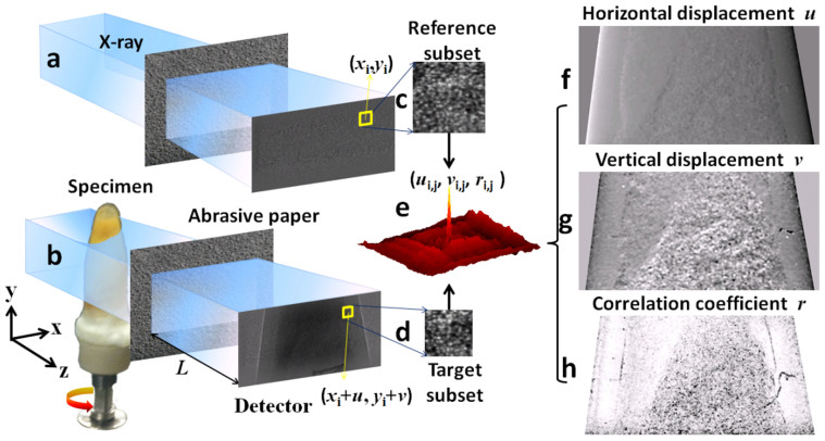
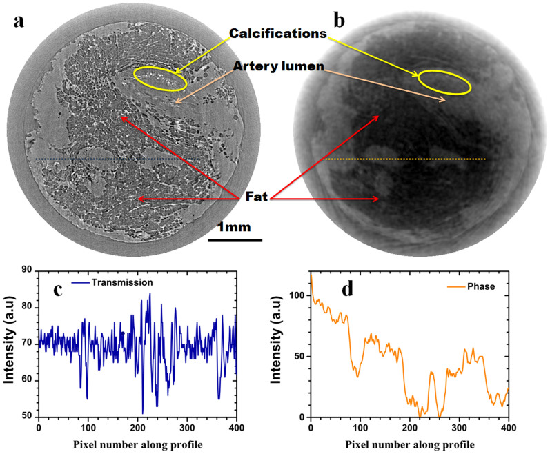
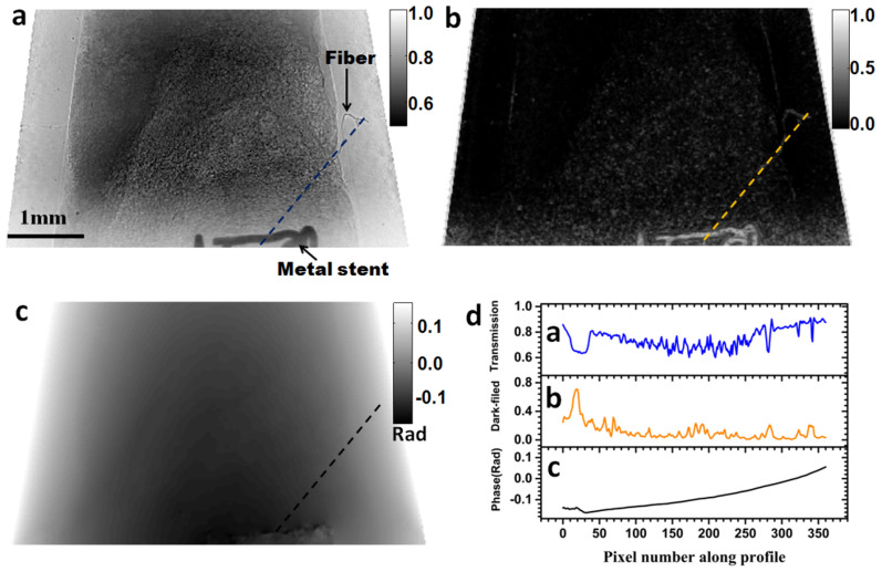
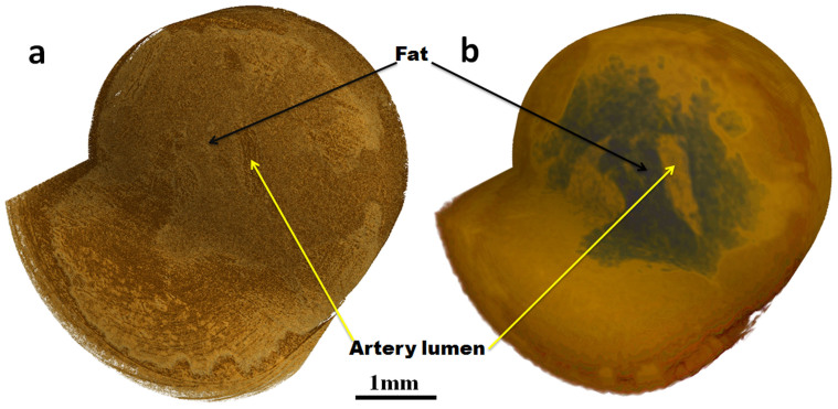
X-ray imaging techniques that capture variations in the x-ray phase can yield higher contrast images with lower x-ray dose than is possible with conventional absorption radiography. However, the extraction of phase information is often more difficult than the extraction of absorption information and requires a more sophisticated experimental arrangement. We here report a method for three-dimensional (3D) X-ray phase contrast computed tomography (CT) which gives quantitative volumetric information on the real part of the refractive index. The method is based on the recently developed X-ray speckle tracking technique in which the displacement of near field speckle is tracked using a digital image correlation algorithm. In addition to differential phase contrast projection images, the method allows the dark-field images to be simultaneously extracted. After reconstruction, compared to conventional absorption CT images, the 3D phase CT images show greatly enhanced contrast. This new imaging method has advantages compared to other X-ray imaging methods in simplicity of experimental arrangement, speed of measurement and relative insensitivity to beam movements. These features make the technique an attractive candidate for material imaging such as in-vivo imaging of biological systems containing soft tissue.

摘要

能够捕捉X射线相位变化的X射线成像技术,与传统吸收式射线照相相比,能够以更低的X射线剂量产生对比度更高的图像。然而,相位信息的提取通常比吸收信息的提取更困难,并且需要更复杂的实验装置。我们在此报告一种用于三维(3D)X射线相衬计算机断层扫描(CT)的方法,该方法可提供关于折射率实部的定量体积信息。该方法基于最近开发的X射线散斑跟踪技术,其中使用数字图像相关算法跟踪近场散斑的位移。除了微分相衬投影图像外,该方法还允许同时提取暗场图像。重建后,与传统吸收式CT图像相比,3D相衬CT图像显示出对比度大大增强。与其他X射线成像方法相比,这种新的成像方法在实验装置的简单性、测量速度以及对光束移动的相对不敏感性方面具有优势。这些特性使该技术成为材料成像的有吸引力的候选方法,例如对包含软组织的生物系统进行体内成像。

https://cdn.ncbi.nlm.nih.gov/pmc/blobs/4c33/4349152/15acaa36fd55/srep08762-f4.jpg
https://cdn.ncbi.nlm.nih.gov/pmc/blobs/4c33/4349152/f08a17117bb7/srep08762-f1.jpg
https://cdn.ncbi.nlm.nih.gov/pmc/blobs/4c33/4349152/6d6d2e04604f/srep08762-f2.jpg
https://cdn.ncbi.nlm.nih.gov/pmc/blobs/4c33/4349152/4f3609be21e0/srep08762-f3.jpg
https://cdn.ncbi.nlm.nih.gov/pmc/blobs/4c33/4349152/15acaa36fd55/srep08762-f4.jpg

相似文献

1
X-ray phase contrast tomography by tracking near field speckle.通过跟踪近场散斑进行X射线相衬断层扫描
Sci Rep. 2015 Mar 4;5:8762. doi: 10.1038/srep08762.
2
A new method to retrieve phase information for equiangular fan beam differential phase contrast computed tomography.一种用于等角扇束差分相位对比计算机断层成像的相位信息提取新方法。
Med Phys. 2013 Mar;40(3):031911. doi: 10.1118/1.4791672.
3
Cardiac motion correction based on partial angle reconstructed images in x-ray CT.基于X射线CT部分角度重建图像的心脏运动校正
Med Phys. 2015 May;42(5):2560-71. doi: 10.1118/1.4918580.
4
A micro-tomography method based on X-ray diffraction enhanced imaging for the visualization of micro-organs and soft tissues.一种基于X射线衍射增强成像的显微断层扫描方法,用于微器官和软组织的可视化。
Comput Med Imaging Graph. 2006 Sep-Oct;30(6-7):339-47. doi: 10.1016/j.compmedimag.2006.09.003. Epub 2006 Oct 18.
5
[Two-dimensional Projection and Image Reconstruction].[二维投影与图像重建]
Igaku Butsuri. 2015;35(3):217-22.
6
Characteristics of a newly developed dentomaxillofacial X-ray cone beam CT scanner (CB MercuRay): system configuration and physical properties.新开发的口腔颌面X射线锥形束CT扫描仪(CB MercuRay)的特点:系统配置和物理特性。
Dentomaxillofac Radiol. 2004 Jan;33(1):51-9. doi: 10.1259/dmfr/54013049.
7
Optimisation of image reconstruction for phase-contrast x-ray Talbot-Lau imaging with regard to mechanical robustness.关于机械稳健性的相衬X射线塔尔博特-劳成像的图像重建优化
Phys Med Biol. 2016 Sep 7;61(17):6441-64. doi: 10.1088/0031-9155/61/17/6441. Epub 2016 Aug 12.
8
Three-dimensional reconstruction of teeth and jaws based on segmentation of CT images using watershed transformation.基于分水岭变换的CT图像分割的牙齿和颌骨三维重建。
Dentomaxillofac Radiol. 2015;44(4):20140313. doi: 10.1259/dmfr.20140313. Epub 2015 Jan 7.
9
Statistical shape model-based reconstruction of a scaled, patient-specific surface model of the pelvis from a single standard AP x-ray radiograph.基于统计形状模型的骨盆单张标准前后位 X 射线片重建患者个体化缩放表面模型。
Med Phys. 2010 Apr;37(4):1424-39. doi: 10.1118/1.3327453.
10
Linac-integrated 4D cone beam CT: first experimental results.直线加速器集成式4D锥形束CT:首次实验结果。
Phys Med Biol. 2006 Jun 7;51(11):2939-52. doi: 10.1088/0031-9155/51/11/017. Epub 2006 May 24.

引用本文的文献

1
Development of a nanometre scale X-ray speckle-based CT technique through the 3-D histological assessment of an acute respiratory distress syndrome model.基于纳米级 X 射线散斑的 CT 技术的发展,通过急性呼吸窘迫综合征模型的 3D 组织学评估。
Sci Rep. 2024 Oct 10;14(1):23745. doi: 10.1038/s41598-024-72660-0.
2
Dynamic X-ray speckle-tracking imaging with high-accuracy phase retrieval based on deep learning.基于深度学习的高精度相位恢复动态X射线散斑跟踪成像
IUCrJ. 2024 Jan 1;11(Pt 1):73-81. doi: 10.1107/S2052252523010114.
3
Two-dimensional speckle technique for slope error measurements of weakly focusing reflective X-ray optics.

本文引用的文献

1
Speckle-based x-ray phase-contrast and dark-field imaging with a laboratory source.基于散斑的 X 射线相衬和暗场成像与实验室光源。
Phys Rev Lett. 2014 Jun 27;112(25):253903. doi: 10.1103/PhysRevLett.112.253903. Epub 2014 Jun 26.
2
A sensitive x-ray phase contrast technique for rapid imaging using a single phase grid analyzer.一种使用单个相栅分析器进行快速成像的灵敏 X 射线相衬技术。
Opt Lett. 2013 Nov 15;38(22):4605-8. doi: 10.1364/OL.38.004605.
3
Translation of atherosclerotic plaque phase-contrast CT imaging from synchrotron radiation to a conventional lab-based X-ray source.
用于弱聚焦反射式X射线光学元件斜率误差测量的二维散斑技术。
J Synchrotron Radiat. 2022 Nov 1;29(Pt 6):1385-1393. doi: 10.1107/S160057752200916X. Epub 2022 Oct 5.
4
High-Resolution Scanning Coded-Mask-Based X-ray Multi-Contrast Imaging and Tomography.基于高分辨率扫描编码掩模的X射线多对比度成像与断层扫描
J Imaging. 2021 Nov 24;7(12):249. doi: 10.3390/jimaging7120249.
5
Nano-precision metrology of X-ray mirrors with laser speckle angular measurement.基于激光散斑角度测量的X射线镜纳米精度计量学
Light Sci Appl. 2021 Sep 22;10(1):195. doi: 10.1038/s41377-021-00632-4.
6
Quantitative Phase and Intensity Microscopy Using Snapshot White Light Wavefront Sensing.利用快照白光波前传感的定量相位和强度显微镜。
Sci Rep. 2019 Sep 24;9(1):13795. doi: 10.1038/s41598-019-50264-3.
7
High-energy, high-resolution, fly-scan X-ray phase tomography.高能量、高分辨率、飞扫 X 射线相衬断层成像。
Sci Rep. 2019 Jun 20;9(1):8913. doi: 10.1038/s41598-019-45561-w.
8
Fast X-ray Differential Phase Contrast Imaging with One Exposure and without Movements.单次曝光无运动的快速 X 射线差分相位衬度成像。
Sci Rep. 2019 Feb 4;9(1):1113. doi: 10.1038/s41598-018-37687-0.
9
X-ray phase-contrast imaging with engineered porous materials over 50 keV.使用能量超过50千电子伏特的工程多孔材料进行的X射线相衬成像。
J Synchrotron Radiat. 2018 Jul 1;25(Pt 4):1182-1188. doi: 10.1107/S1600577518005623. Epub 2018 Jun 12.
10
Periodic microstructures of blood capillaries revealed by synchrotron X-ray multi-resolution microscopic analysis.同步加速器X射线多分辨率显微镜分析揭示的毛细血管周期性微观结构
Biomed Opt Express. 2017 Nov 30;8(12):5825-5833. doi: 10.1364/BOE.8.005825. eCollection 2017 Dec 1.
从同步辐射到常规实验室基于 X 射线源的动脉粥样硬化斑块相衬 CT 成像的转换。
PLoS One. 2013 Sep 9;8(9):e73513. doi: 10.1371/journal.pone.0073513. eCollection 2013.
4
Two-dimensional x-ray beam phase sensing.二维 X 射线光束相位传感。
Phys Rev Lett. 2012 Apr 13;108(15):158102. doi: 10.1103/PhysRevLett.108.158102. Epub 2012 Apr 11.
5
Two-dimensional grating-based X-ray phase-contrast imaging using Fourier transform phase retrieval.基于二维光栅的X射线相衬成像:傅里叶变换相位恢复法
Opt Express. 2011 Feb 14;19(4):3339-46. doi: 10.1364/OE.19.003339.
6
Single-shot x-ray differential phase-contrast and diffraction imaging using two-dimensional transmission gratings.基于二维透射光栅的单次 X 射线差分相位衬度和衍射成像。
Opt Lett. 2010 Jun 15;35(12):1932-4. doi: 10.1364/OL.35.001932.
7
Directional x-ray dark-field imaging.定向 X 射线暗场成像。
Phys Med Biol. 2010 Jun 21;55(12):3317-23. doi: 10.1088/0031-9155/55/12/004. Epub 2010 May 18.
8
High-speed X-ray phase imaging and X-ray phase tomography with Talbot interferometer and white synchrotron radiation.利用塔尔博特干涉仪和白色同步辐射进行高速X射线相成像和X射线相断层扫描。
Opt Express. 2009 Jul 20;17(15):12540-5. doi: 10.1364/oe.17.012540.
9
A two-directional approach for grating based differential phase contrast imaging using hard x-rays.一种基于光栅的硬X射线差分相衬成像的双向方法。
Opt Express. 2007 Feb 5;15(3):1175-81. doi: 10.1364/oe.15.001175.
10
X-ray phase imaging with a grating interferometer.使用光栅干涉仪的X射线相成像
Opt Express. 2005 Aug 8;13(16):6296-304. doi: 10.1364/opex.13.006296.