Ulrich Xialing, Yablonskiy Dmitriy A
Department of Radiology, Washington University, St. Louis, Missouri, USA.
Magn Reson Med. 2016 Feb;75(2):606-15. doi: 10.1002/mrm.25610. Epub 2015 Mar 10.
The development of a reliable clinical technique for quantitative measurements of the parameters defining the BOLD effect, i.e., oxygen extraction fraction (OEF), and deoxygenated cerebral blood volume, dCBV, is needed to study brain function in health and disease. Herein we propose such a technique that is based on a widely available gradient recalled echo (GRE) MRI.
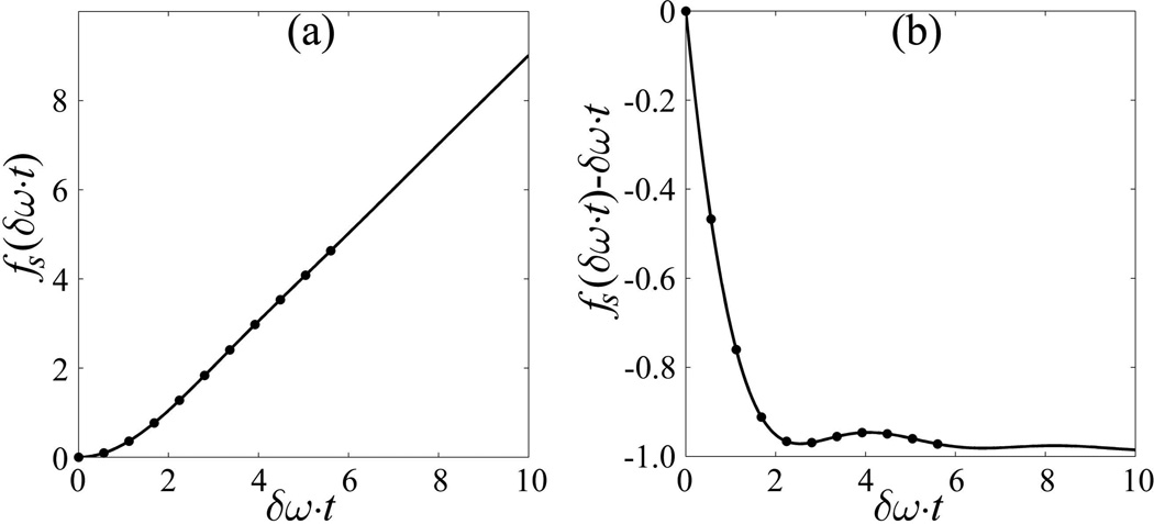
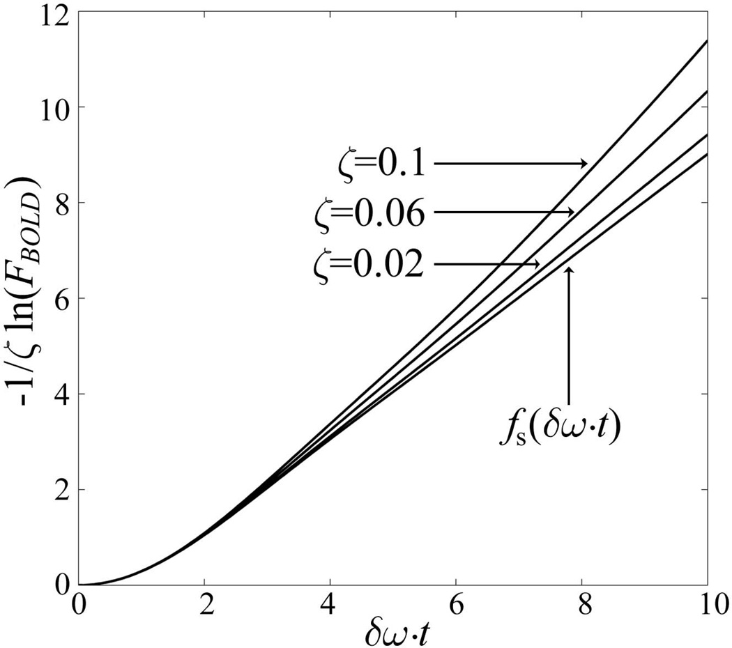
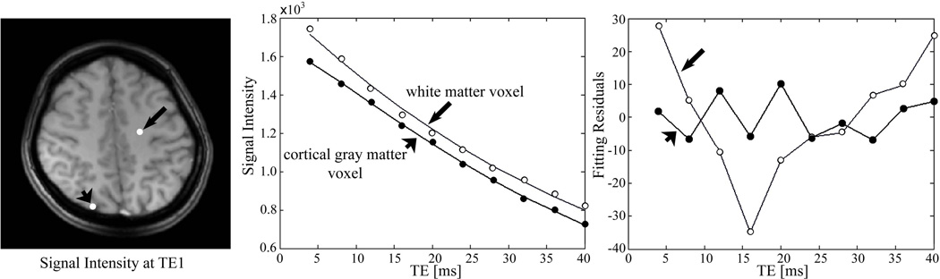
Our method is based on GRE with multiple echoes and a model of signal decay (Yablonskiy, MRM 1998) that takes into account microscopic cellular (R2), mesoscopic (BOLD), and macroscopic (background field gradients) contributions to the GRE signal decay with additional accounting for physiologic fluctuations.
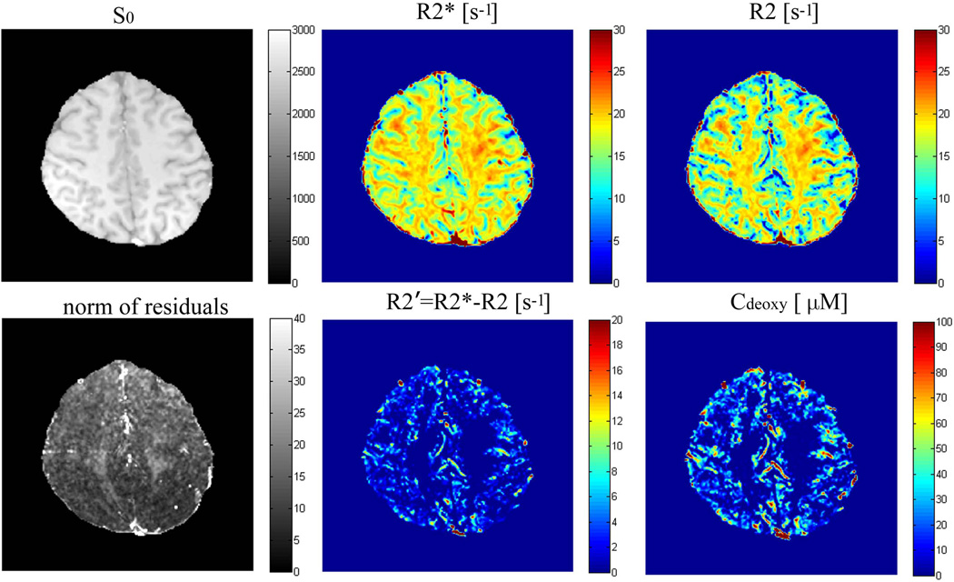
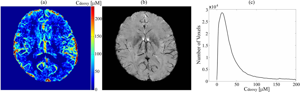
Using 3 Tesla MRI, we generate high resolution quantitative maps of R2*, R2, R2', and tissue concentration of deoxyhemoglobin, the latter providing a quantitative version of SWI. Our results for OEF and dCBV in gray matter are in a reasonable agreement with the literature data.
The proposed approach allows generating high resolution maps of hemodynamic parameters using clinical MRI. The technique can be applied to study such tissues as gray matter, tumors, etc.; however, it requires further development for use in tissues where extra- and intracellular compartments possess substantially different frequencies and relaxation properties (e.g., white matter).
为了研究健康和疾病状态下的脑功能,需要开发一种可靠的临床技术,用于定量测量定义血氧水平依赖(BOLD)效应的参数,即氧提取分数(OEF)和脱氧脑血容量(dCBV)。在此,我们提出一种基于广泛可用的梯度回波(GRE)MRI的技术。
我们的方法基于具有多个回波的GRE以及信号衰减模型(Yablonskiy,《磁共振成像杂志》1998年),该模型考虑了微观细胞(R2)、介观(BOLD)和宏观(背景场梯度)对GRE信号衰减的贡献,并额外考虑了生理波动。
使用3特斯拉MRI,我们生成了R2*、R2、R2'和脱氧血红蛋白组织浓度的高分辨率定量图,后者提供了磁敏感加权成像(SWI)的定量版本。我们在灰质中获得的OEF和dCBV结果与文献数据合理一致。
所提出的方法允许使用临床MRI生成血流动力学参数的高分辨率图。该技术可应用于研究灰质、肿瘤等组织;然而,对于细胞外和细胞内隔室具有显著不同频率和弛豫特性的组织(如白质),还需要进一步改进才能使用。