Bosco Alejandra, Romero Cesar O, Breen Kevin T, Chagovetz Alexis A, Steele Michael R, Ambati Balamurali K, Vetter Monica L
Department of Neurobiology and Anatomy, University of Utah, Salt Lake City, UT 84132, USA
Department of Neurobiology and Anatomy, University of Utah, Salt Lake City, UT 84132, USA.
Dis Model Mech. 2015 May;8(5):443-55. doi: 10.1242/dmm.018788. Epub 2015 Mar 9.
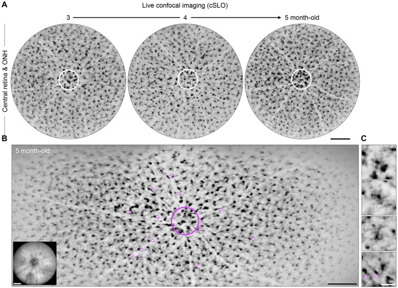
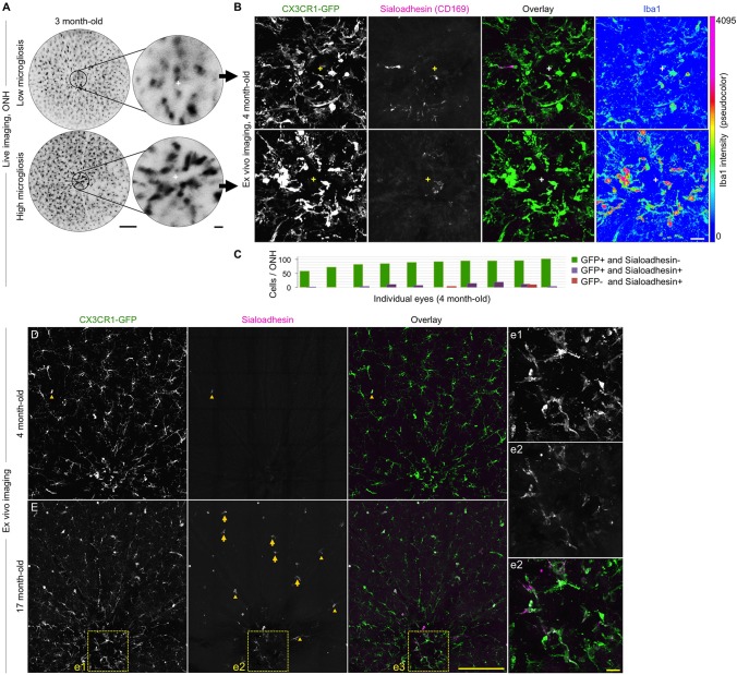
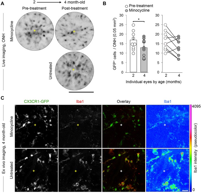
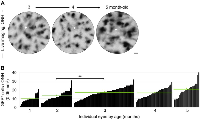
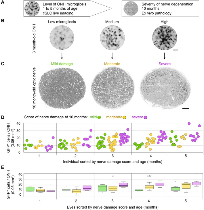
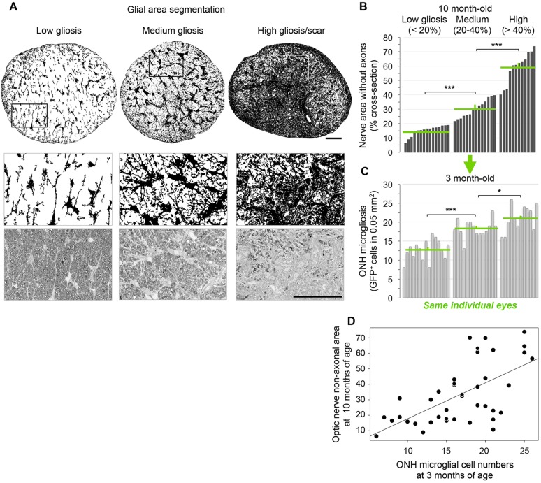
Microglia serve key homeostatic roles, and respond to neuronal perturbation and decline with a high spatiotemporal resolution. The course of all chronic CNS pathologies is thus paralleled by local microgliosis and microglia activation, which begin at early stages of the disease. However, the possibility of using live monitoring of microglia during early disease progression to predict the severity of neurodegeneration has not been explored. Because the retina allows live tracking of fluorescent microglia in their intact niche, here we investigated their early changes in relation to later optic nerve neurodegeneration. To achieve this, we used the DBA/2J mouse model of inherited glaucoma, which develops progressive retinal ganglion cell degeneration of variable severity during aging, and represents a useful model to study pathogenic mechanisms of retinal ganglion cell decline that are similar to those in human glaucoma. We imaged CX3CR1(+/GFP) microglial cells in vivo at ages ranging from 1 to 5 months by confocal scanning laser ophthalmoscopy (cSLO) and quantified cell density and morphological activation. We detected early microgliosis at the optic nerve head (ONH), where axonopathy first manifests, and could track attenuation of this microgliosis induced by minocycline. We also observed heterogeneous and dynamic patterns of early microglia activation in the retina. When the same animals were aged and analyzed for the severity of optic nerve pathology at 10 months of age, we found a strong correlation with the levels of ONH microgliosis at 3 to 4 months. Our findings indicate that live imaging and monitoring the time course and levels of early retinal microgliosis and microglia activation in glaucoma could serve as indicators of future neurodegeneration severity.
小胶质细胞发挥着关键的稳态作用,并以高时空分辨率对神经元扰动和衰退做出反应。因此,所有慢性中枢神经系统疾病的病程都伴随着局部小胶质细胞增生和小胶质细胞激活,这在疾病的早期阶段就开始了。然而,在疾病早期进展过程中对小胶质细胞进行实时监测以预测神经退行性变严重程度的可能性尚未得到探索。由于视网膜允许在其完整的微环境中对荧光小胶质细胞进行实时追踪,因此我们在此研究了它们与后期视神经神经退行性变相关的早期变化。为了实现这一目标,我们使用了遗传性青光眼的DBA/2J小鼠模型,该模型在衰老过程中会发生严重程度各异的进行性视网膜神经节细胞变性,是研究与人类青光眼相似的视网膜神经节细胞衰退致病机制的有用模型。我们通过共聚焦扫描激光检眼镜(cSLO)在1至5个月龄的体内对CX3CR1(+/GFP)小胶质细胞进行成像,并量化细胞密度和形态激活。我们在视神经乳头(ONH)检测到早期小胶质细胞增生,轴突病变首先在那里表现出来,并且可以追踪米诺环素诱导的这种小胶质细胞增生的减弱。我们还观察到视网膜中早期小胶质细胞激活的异质性和动态模式。当对相同的动物进行衰老处理并在10个月龄时分析视神经病理的严重程度时,我们发现与3至4个月时ONH小胶质细胞增生的水平有很强的相关性。我们的研究结果表明,在青光眼中对早期视网膜小胶质细胞增生和小胶质细胞激活的时间进程和水平进行实时成像和监测,可以作为未来神经退行性变严重程度的指标。