Dong Wei, Yuwen Zhang, Xiaohui Gong
Department of pediatrics, Tongji University.
Department of Neonatology, Shanghai Children's Hospital, Shanghai Jiao Tong University, Shanghai, China.
Iran J Pediatr. 2014 Aug;24(4):435-40. Epub 2014 Jul 27.
It has been found that asphyxia influences proliferation and differentiation of brain neural stem cells in newborn animal models, and that peripheral blood stem cells play an important role in repairing brain damage. But it has not been reported yet whether asphyxia influences peripheral blood stem cells differentiating into neural cells, and whether with the progress of the disease there is a change of peripheral blood stem cells differentiating into neural cells in newborns with hypoxic ischemic encephalopathy (HIE).
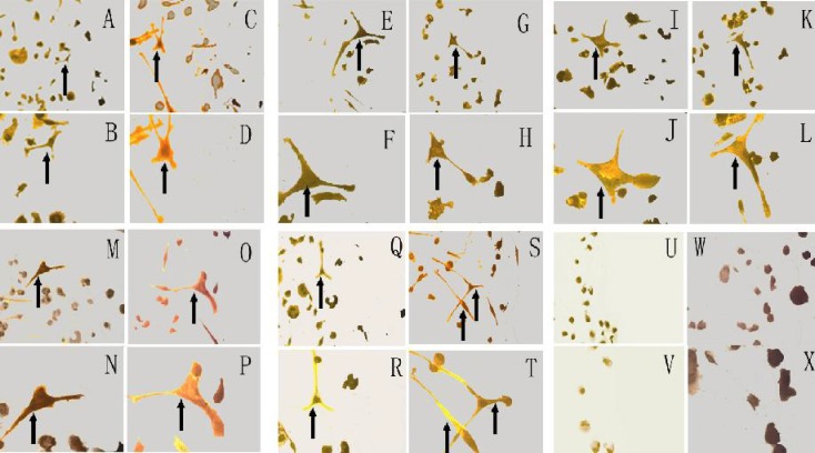
Fifty term HIE infants were enrolled in research from March, 2007 to March, 2010. There were 10 cases of the severe HIE patients with good improvement, the severe HIE patients with poor improvement, the moderate HIE patients, the mild HIE patients and the controls, respectively. The peripheral mononuclear cells collected within 24 hours and on 7th day after birth were cultured in vitro for 10 days to differentiate into neural cells. The induced nestin positive cells were identified with Immunohistochemistry and counted. Findings : Within 24 hours after birth, there were no difference of induced nestin positive cells among the severe HIE patients with good improvement (68.99±7.85), the severe HIE patients with poor improvement (71.43±6.88), the moderate HIE patients (73.34±6.46), the mild HIE patients (70.46±6.66) and the controls (71.13±7.19, F=0.51, P=0.7). In the severe HIE patients with obvious improvement, the induced nestin positive cells from 7th day peripheral blood mononuclear cells (94.50±15.57) increased markedly compared with that within 24 hours (68.99±7.85, t=4.66, P<0.001), and were higher than the induced nestin positive cells from 7(th) day peripheral blood mononuclear cells in the severe HIE patients with no obvious improvement (94.50±15.57 vs 69.48±5.32, t=4.62, P<0.001).
The ability of peripheral mononuclear cells differentiating into neural cells in term infants with good improvement suffering from severe HIE was enhanced, which may suggest possible relationship between the brain repair and the peripheral stem cells.
在新生动物模型中发现,窒息会影响脑内神经干细胞的增殖和分化,并且外周血干细胞在脑损伤修复中发挥重要作用。但窒息是否影响外周血干细胞向神经细胞分化,以及缺氧缺血性脑病(HIE)新生儿外周血干细胞向神经细胞分化是否随病情进展而发生变化,目前尚未见报道。
选取2007年3月至2010年3月期间收治的50例足月HIE患儿,分为重度HIE好转良好组、重度HIE好转不良组、中度HIE组、轻度HIE组及对照组,每组各10例。收集出生后24小时内及出生后第7天的外周血单个核细胞,体外培养10天诱导其分化为神经细胞,采用免疫组织化学法鉴定并计数诱导后巢蛋白阳性细胞。结果:出生后24小时内,重度HIE好转良好组(68.99±7.85)、重度HIE好转不良组(71.43±6.88)、中度HIE组(73.34±6.46)、轻度HIE组(70.46±6.66)及对照组(71.13±7.19)诱导后巢蛋白阳性细胞数比较,差异无统计学意义(F=0.51,P=0.7)。重度HIE好转明显组出生后第7天外周血单个核细胞诱导后巢蛋白阳性细胞数(94.50±15.57)较出生后24小时(68.99±7.85)明显增加(t=4.66,P<0.001),且高于重度HIE好转不明显组出生后第7天外周血单个核细胞诱导后巢蛋白阳性细胞数(94.50±15.57比69.48±5.32,t=4.62,P<0.001)。
重度HIE好转良好的足月新生儿外周血单个核细胞向神经细胞分化的能力增强,提示脑修复与外周干细胞之间可能存在联系。