Farivar Shirin, Mohamadzade Zahra, Shiari Reza, Fahimzad Alireza
Department of Genetics, Faculty of Biological Sciences, Shahid Beheshti University, Tehran, Iran ; Laser and Plasma Research Institute, Shahid Beheshti University, Tehran, Iran.
Department of Genetics, Faculty of Biological Sciences, Shahid Beheshti University, Tehran, Iran.
Iran J Child Neurol. 2015 Winter;9(1):87-93.
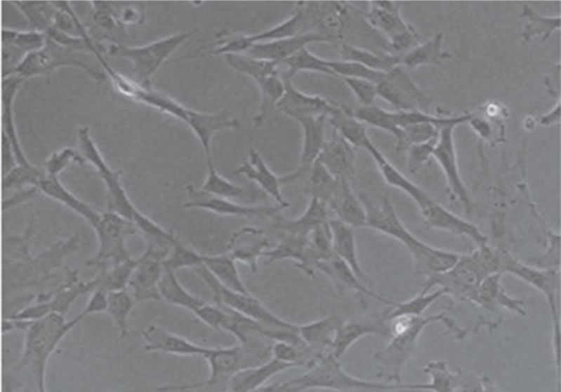
Wharton's jelly (WJ) is the gelatinous connective tissue from the umbilical cord. It is composed of mesenchymal stem cells, collagen fibers, and proteoglycans. The stem cells in WJ have properties that are interesting for research. For example, they are simple to harvest by noninvasive methods, provide large numbers of cells without risk to the donor, the stem cell population may be expanded in vitro, cryogenically stored, thawed, genetically manipulated, and differentiated in vitro. In our study, we investigated the effect of human cerebrospinal fluid (CSF) on neural differentiation of human WJ stem cells.
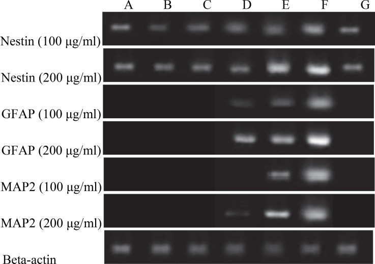
MATERIAL & METHODS: The cells in passage 2 were induced into neural differentiation with different concentrations of human cerebrospinal fluid. Differentiation along with neural lineage was documented by expression of three neural markers: Nestin, Microtubule-Associated Protein 2 (MAP2), and Glial Fibrillary Astrocytic Protein (GFAP) for 21 days. The expression of the identified genes was confirmed by Reverse Transcriptase PCR (RT-PCR).
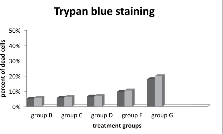
Treatment with 100 and 200μg/ml CSF resulted in the expression of GFAP and glial cells marker on days 14 and 21. The expression of neural-specific genes following CSF treatment was dose-dependent and time-dependent. Treatment of the cells with a twofold concentration of CSF, led to the expression of MAP2 on day 14 of induction. No expression of GFAP was detected before day 14 or MAP2 before day 21, which shows the importance of the treatment period. In the present study, expression analysis for the known neural markers: Nestin, GFAP, and MAP2 using RT-PCR were performed. The data demonstrated that CSF could play a role as a strong inducer.
RT-PCR showed that cerebrospinal fluid promotes the expression of Nestin, MAP2, and GFAP mRNA in a dose-dependent manner, especially at a concentration of 200 μl/ml. In summary, CSF induces neurogenesis of WJ stem cells that encourages tissue engineering applications with these cells for treatments of neurodegenerative defects and traumatic brain injury.
华通氏胶(WJ)是脐带中的凝胶状结缔组织。它由间充质干细胞、胶原纤维和蛋白聚糖组成。WJ中的干细胞具有一些值得研究的特性。例如,它们可以通过非侵入性方法轻松获取,能提供大量细胞且对供体无风险,干细胞群体可在体外扩增、低温保存、解冻、进行基因操作并在体外分化。在我们的研究中,我们调查了人脑脊液(CSF)对人WJ干细胞神经分化的影响。
用不同浓度的人脑脊液诱导第2代细胞进行神经分化。通过三种神经标志物:巢蛋白、微管相关蛋白2(MAP2)和胶质纤维酸性蛋白(GFAP)的表达记录21天内的神经谱系分化情况。通过逆转录聚合酶链反应(RT-PCR)确认已鉴定基因的表达。
用100和200μg/ml脑脊液处理导致在第14天和第21天GFAP和胶质细胞标志物的表达。脑脊液处理后神经特异性基因的表达呈剂量依赖性和时间依赖性。用两倍浓度的脑脊液处理细胞,导致在诱导第14天MAP2的表达。在第14天之前未检测到GFAP的表达,在第21天之前未检测到MAP2的表达,这表明处理时间的重要性。在本研究中,使用RT-PCR对已知神经标志物:巢蛋白、GFAP和MAP2进行了表达分析。数据表明脑脊液可作为一种强大的诱导剂发挥作用。
RT-PCR表明脑脊液以剂量依赖性方式促进巢蛋白、MAP2和GFAP mRNA的表达,尤其是在浓度为200μl/ml时。总之,脑脊液诱导WJ干细胞的神经发生,这鼓励了利用这些细胞进行组织工程应用以治疗神经退行性缺陷和创伤性脑损伤。