Butler Holly J, Fogarty Simon W, Kerns Jemma G, Martin-Hirsch Pierre L, Fullwood Nigel J, Martin Francis L
Centre for Biophotonics, Lancaster Environment Centre, Lancaster University, Bailrigg, Lancaster LA1 4YQ, UK.
Analyst. 2015 May 7;140(9):3090-7. doi: 10.1039/c4an01899k. Epub 2015 Mar 24.
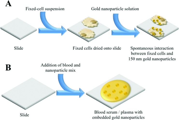
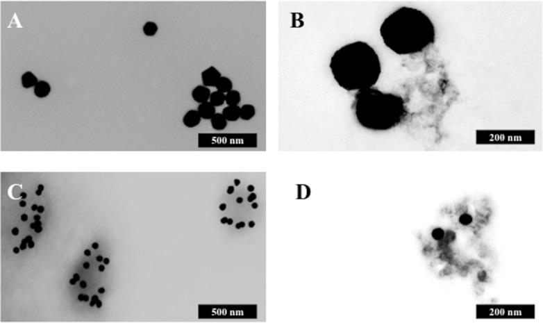
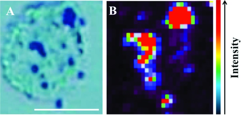
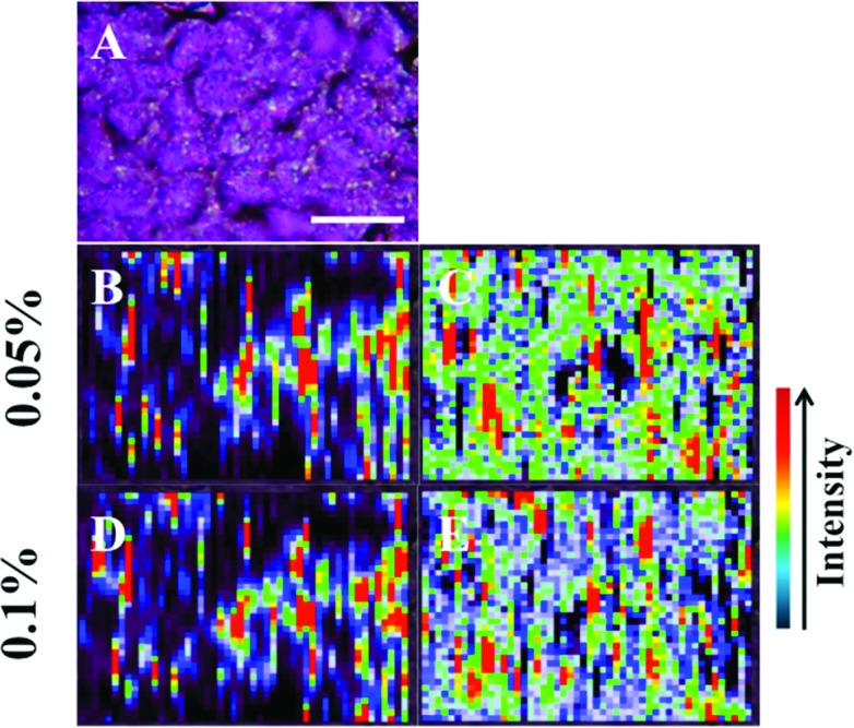
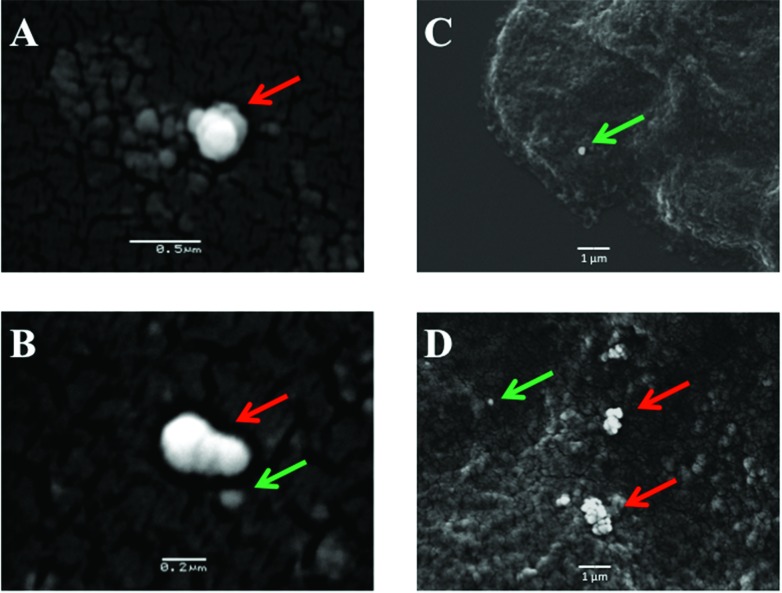
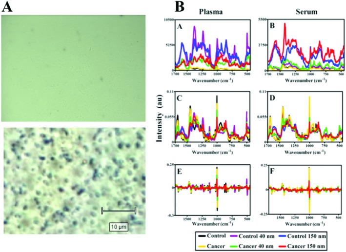
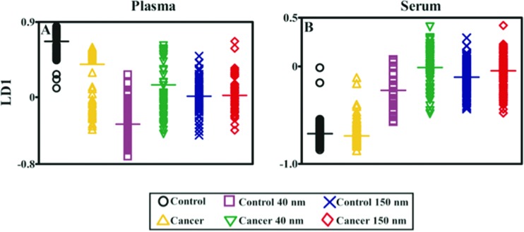
As biospectroscopy techniques continue to be developed for screening or diagnosis within a point-of-care setting, an important development for this field will be high-throughput optimization. For many of these techniques, it is therefore necessary to adapt and develop parameters to generate a robust yet simple approach delivering high-quality spectra from biological samples. Specifically, this is important for surface-enhanced Raman spectroscopy (SERS) wherein there are multiple variables that can be optimised to achieve an enhancement of the Raman signal from a sample. One hypothesis is that "large" diameter (>100 nm) gold nanoparticles provide a greater enhancement at near-infrared (NIR) and infrared (IR) wavelengths than those <100 nm in diameter. Herein, we examine this notion using examples in which SERS spectra were acquired from MCF-7 breast cancer cells incubated with 150 nm gold nanoparticles. It was found that 150 nm gold nanoparticles are an excellent material for NIR/IR SERS. Larger gold nanoparticles may better satisfy the theoretical restraints for SERS enhancement at NIR/IR wavelengths compared to smaller nanoparticles. Also, larger nanoparticles or their aggregates are more readily observed via optical microscopy (and especially electron microscopy) compared to smaller ones. This allows rapid and straightforward identification of target areas containing a high concentration of nanoparticles and facilitating SERS spectral acquisition. To some extent, these observations appear to extend to biofluids such as blood plasma or (especially) serum; SERS spectra of such biological samples often exhibit a low signal-to-noise ratio in the absence of nanoparticles. With protein-rich biofluids such as serum, a dramatic SERS effect can be observed; although this might facilitate improved spectral biomarker identification in the future, it may not always improve classification between control vs. cancer. Thus, use of "large" gold nanoparticles are a good starting point in order to derive informative NIR/IR SERS analysis of biological samples.
随着生物光谱技术不断发展以用于即时护理环境中的筛查或诊断,该领域的一项重要进展将是高通量优化。因此,对于许多此类技术而言,有必要调整和开发参数,以生成一种稳健且简单的方法,从生物样品中获取高质量光谱。具体而言,这对于表面增强拉曼光谱(SERS)很重要,因为在SERS中有多个变量可以优化,以实现样品拉曼信号的增强。一种假设是,直径“大”(>100 nm)的金纳米颗粒在近红外(NIR)和红外(IR)波长下比直径<100 nm的金纳米颗粒具有更大的增强效果。在此,我们通过从与150 nm金纳米颗粒孵育的MCF-7乳腺癌细胞中获取SERS光谱的示例来检验这一概念。结果发现,150 nm金纳米颗粒是用于近红外/红外SERS的优良材料。与较小的纳米颗粒相比,较大的金纳米颗粒可能更能满足近红外/红外波长下SERS增强的理论限制。此外,与较小的纳米颗粒相比,较大的纳米颗粒或其聚集体通过光学显微镜(尤其是电子显微镜)更容易观察到。这使得能够快速直接地识别含有高浓度纳米颗粒的目标区域,并便于进行SERS光谱采集。在某种程度上,这些观察结果似乎也适用于生物流体,如血浆或(尤其是)血清;在没有纳米颗粒的情况下,此类生物样品的SERS光谱通常具有较低的信噪比。对于富含蛋白质的生物流体如血清,可以观察到显著的SERS效应;尽管这可能在未来有助于改进光谱生物标志物的识别,但它不一定总能改善对照与癌症之间的分类。因此,使用“大”金纳米颗粒是进行生物样品近红外/红外SERS信息分析的一个良好起点。