Polis Bartosz, Imiela Anna, Polis Lech, Abramczyk Halina
Department of Neurosurgery and Neurotraumatology, Polish Mother's Memorial Hospital Research Institute, 281/289 Rzgowska St., 93-338, Lodz, Poland.
Laboratory of Laser Molecular Spectroscopy, Institute of Applied Radiation Chemistry, Faculty of Chemistry, Lodz University of Technology, Wroblewskiego 15, 93-590, Lodz, Poland.
Childs Nerv Syst. 2018 Dec;34(12):2425-2430. doi: 10.1007/s00381-018-3906-7. Epub 2018 Jul 12.
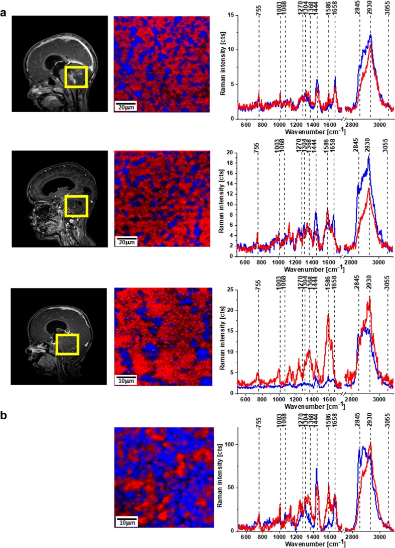
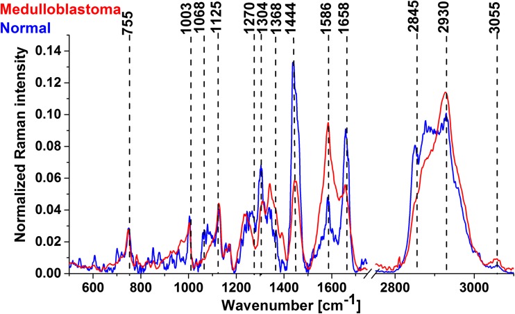
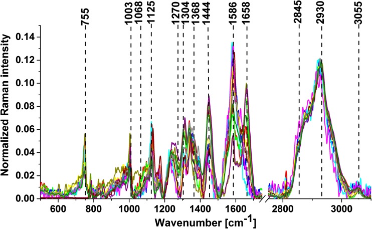
The aim of the study is to use Raman spectroscopy to analyze the biochemical composition of medulloblastoma and normal tissues from the safety margin of the CNS and to find specific Raman biomarkers capable of differentiating between tumorous and normal tissues.
The tissue samples consisted of medulloblastoma (grade IV) (n = 11). The tissues from the negative margins were used as normal controls. Raman images were generated by a confocal Raman microscope-WITec alpha 300 RSA.
Raman vibrational signatures can predict which tissue has tumorous biochemistry and can identify medulloblastoma. The Raman technique makes use of the fact that tumors contain large amounts of protein and far less lipids (fatty compounds), while healthy tissue is rich in both.
The ability of Raman spectroscopy and imaging to detect medulloblastoma tumors fills the niche in diagnostics. These powerful analytical techniques are capable of monitoring tissue morphology and biochemistry. Our results demonstrate that RS can be used to discriminate between normal and medulloblastoma tissues.
本研究旨在利用拉曼光谱分析髓母细胞瘤以及中枢神经系统安全切缘的正常组织的生化组成,并寻找能够区分肿瘤组织和正常组织的特定拉曼生物标志物。
组织样本包括髓母细胞瘤(IV级)(n = 11)。阴性切缘的组织用作正常对照。拉曼图像由共聚焦拉曼显微镜-WITec alpha 300 RSA生成。
拉曼振动特征可以预测哪些组织具有肿瘤生化特性,并能识别髓母细胞瘤。拉曼技术利用了肿瘤含有大量蛋白质而脂质(脂肪化合物)含量远低于正常组织这一事实,而健康组织则富含蛋白质和脂质。
拉曼光谱和成像检测髓母细胞瘤肿瘤的能力填补了诊断领域的空白。这些强大的分析技术能够监测组织形态和生化组成。我们的结果表明拉曼光谱可用于区分正常组织和髓母细胞瘤组织。