• 文献检索
  • 文档翻译
  • 深度研究
  • 学术资讯
  • Suppr Zotero 插件Zotero 插件
  • 邀请有礼
  • 套餐&价格
  • 历史记录
应用&插件
Suppr Zotero 插件Zotero 插件浏览器插件Mac 客户端Windows 客户端微信小程序
定价
高级版会员购买积分包购买API积分包
服务
文献检索文档翻译深度研究API 文档MCP 服务
关于我们
关于 Suppr公司介绍联系我们用户协议隐私条款
关注我们

Suppr 超能文献

核心技术专利:CN118964589B侵权必究
粤ICP备2023148730 号-1Suppr @ 2026

文献检索

告别复杂PubMed语法,用中文像聊天一样搜索,搜遍4000万医学文献。AI智能推荐,让科研检索更轻松。

立即免费搜索

文件翻译

保留排版,准确专业,支持PDF/Word/PPT等文件格式,支持 12+语言互译。

免费翻译文档

深度研究

AI帮你快速写综述,25分钟生成高质量综述,智能提取关键信息,辅助科研写作。

立即免费体验

碲化镉量子点纳米颗粒在哺乳动物肝细胞中的线粒体毒性

Mitochondrial Toxicity of Cadmium Telluride Quantum Dot Nanoparticles in Mammalian Hepatocytes.

作者信息

Nguyen Kathy C, Rippstein Peter, Tayabali Azam F, Willmore William G

机构信息

*Biotechnology Laboratory, Mechanistic Studies Division, Environmental Health Science Research Bureau, HECSB, Health Canada, Ottawa, Ontario, Canada K1A 0K9, Department of Biology and Institute of Biochemistry, Carleton University, Ottawa, Ontario, Canada K1S 5B6 and The University of Ottawa, Heart Institute, Ottawa, Ontario, Canada K1Y 4W7 *Biotechnology Laboratory, Mechanistic Studies Division, Environmental Health Science Research Bureau, HECSB, Health Canada, Ottawa, Ontario, Canada K1A 0K9, Department of Biology and Institute of Biochemistry, Carleton University, Ottawa, Ontario, Canada K1S 5B6 and The University of Ottawa, Heart Institute, Ottawa, Ontario, Canada K1Y 4W7.

*Biotechnology Laboratory, Mechanistic Studies Division, Environmental Health Science Research Bureau, HECSB, Health Canada, Ottawa, Ontario, Canada K1A 0K9, Department of Biology and Institute of Biochemistry, Carleton University, Ottawa, Ontario, Canada K1S 5B6 and The University of Ottawa, Heart Institute, Ottawa, Ontario, Canada K1Y 4W7.

出版信息

Toxicol Sci. 2015 Jul;146(1):31-42. doi: 10.1093/toxsci/kfv068. Epub 2015 Mar 25.

DOI:10.1093/toxsci/kfv068
PMID:25809595
原文链接:https://pmc.ncbi.nlm.nih.gov/articles/PMC4476459/
Abstract

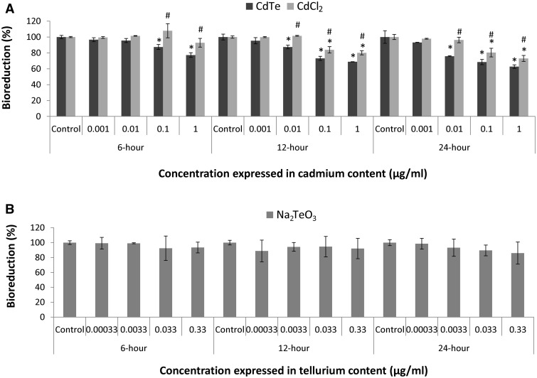
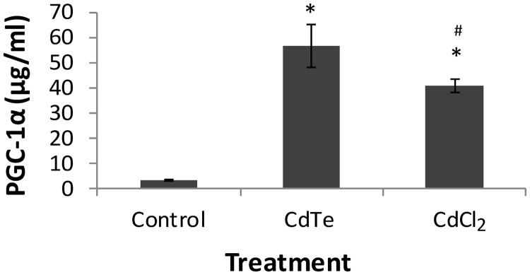
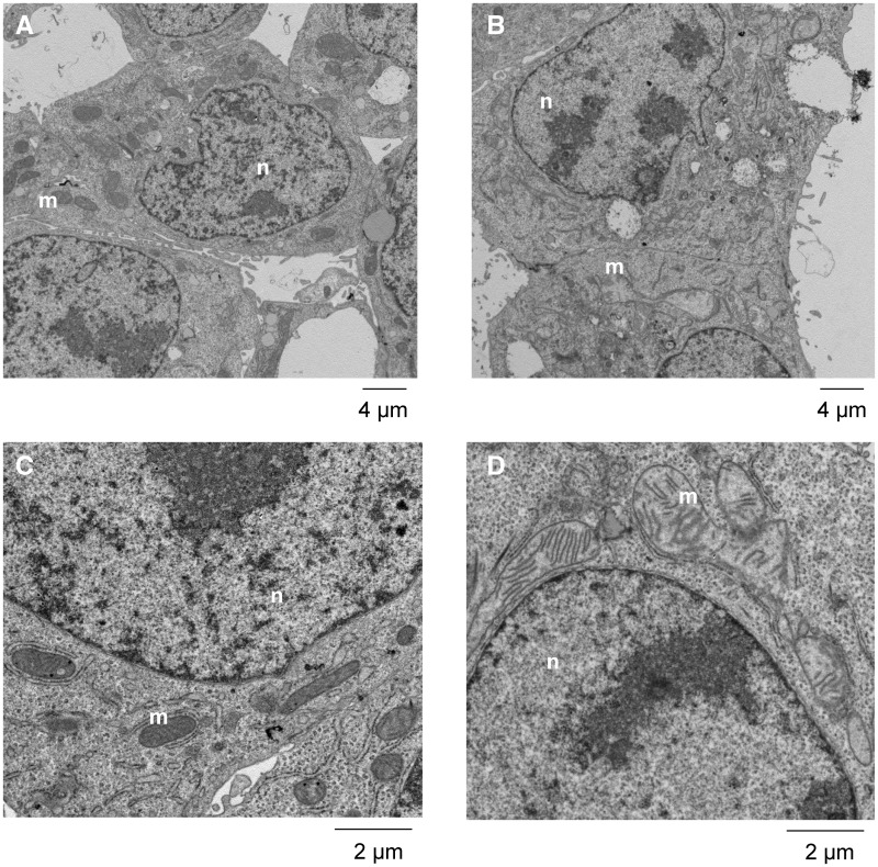
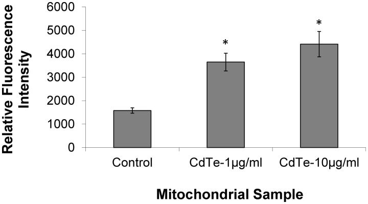
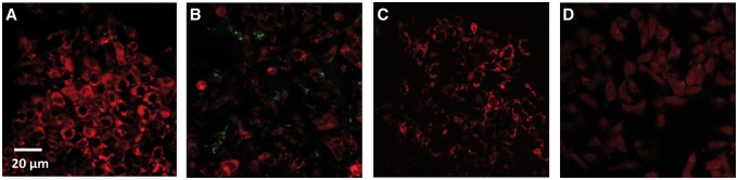
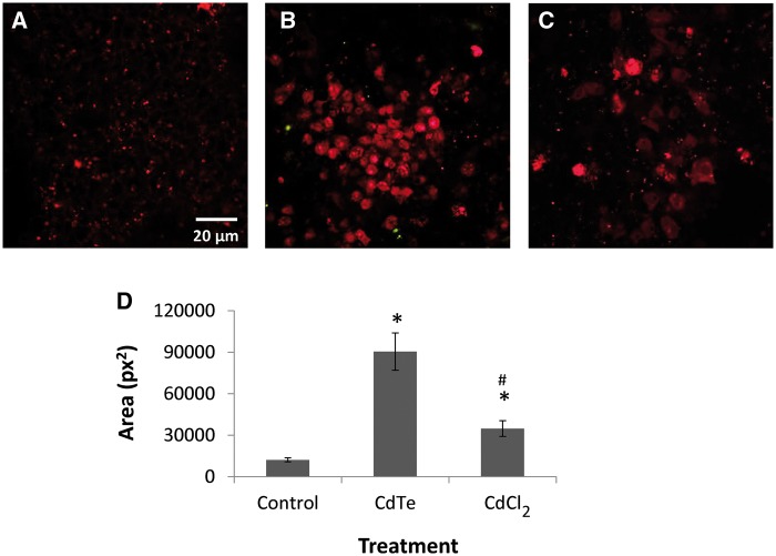
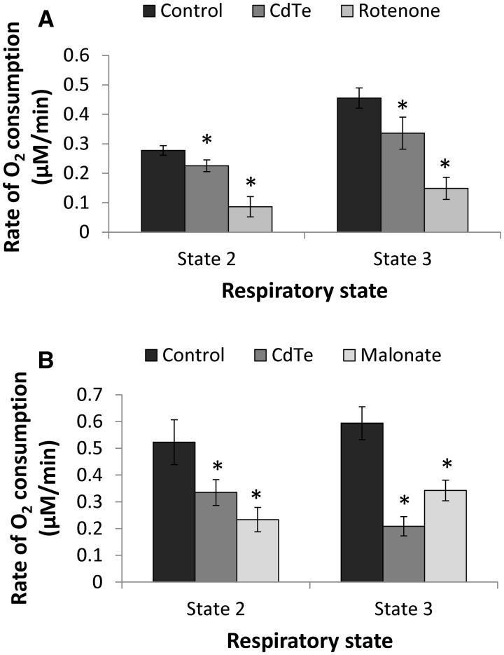
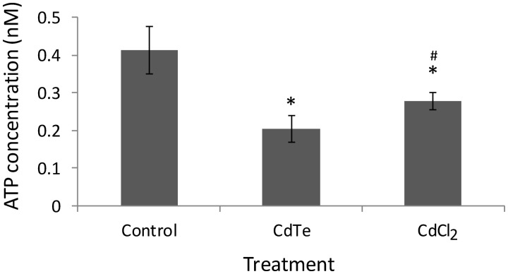
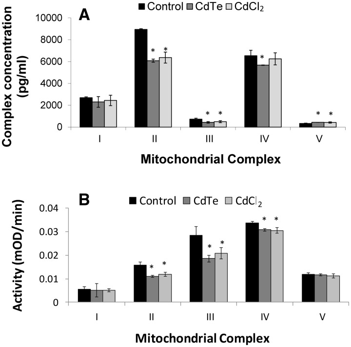
There are an increasing number of studies indicating that mitochondria are relevant targets in nanomaterial-induced toxicity. However, the underlying mechanisms by which nanoparticles (NPs) interact with these organelles and affect their functions are unknown. The aim of this study was to investigate the effects of cadmium telluride quantum dot (CdTe-QD) NPs on mitochondria in human hepatocellular carcinoma HepG2 cells. CdTe-QD treatment resulted in the enlargement of mitochondria as examined with transmission electron microscopy and confocal microscopy. CdTe-QDs appeared to associate with the isolated mitochondria as detected by their inherent fluorescence. Further analyses revealed that CdTe-QD caused disruption of mitochondrial membrane potential, increased intracellular calcium levels, impaired cellular respiration, and decreased adenosine triphosphate synthesis. The effects of CdTe-QDs on mitochondrial oxidative phosphorylation were evidenced by changes in levels and activities of the enzymes of the electron transport chain. Elevation of peroxisome proliferator-activated receptor-γ coactivator levels after CdTe-QD treatment suggested the effects of CdTe-QDs on mitochondrial biogenesis. Our results also showed that the effects of CdTe-QDs were similar or greater to those of cadmium chloride at equivalent concentrations of cadmium, suggesting that the toxic effects of CdTe-QDs were not solely due to cadmium released from the NPs. Overall, the study demonstrated that CdTe-QDs induced multifarious toxicity by causing changes in mitochondrial morphology and structure, as well as impairing their function and stimulating their biogenesis.

摘要

越来越多的研究表明,线粒体是纳米材料诱导毒性的相关靶点。然而,纳米颗粒(NPs)与这些细胞器相互作用并影响其功能的潜在机制尚不清楚。本研究的目的是探讨碲化镉量子点(CdTe-QD)NPs对人肝癌HepG2细胞中线粒体的影响。透射电子显微镜和共聚焦显微镜检查显示,CdTe-QD处理导致线粒体增大。通过其固有荧光检测发现,CdTe-QDs似乎与分离的线粒体相关联。进一步分析表明,CdTe-QD导致线粒体膜电位破坏、细胞内钙水平升高、细胞呼吸受损以及三磷酸腺苷合成减少。电子传递链酶的水平和活性变化证明了CdTe-QDs对线粒体氧化磷酸化的影响。CdTe-QD处理后过氧化物酶体增殖物激活受体-γ共激活因子水平升高,表明CdTe-QDs对线粒体生物发生有影响。我们的结果还表明,在镉浓度相等时,CdTe-QDs的影响与氯化镉相似或更大,这表明CdTe-QDs的毒性作用并非仅归因于从NPs中释放的镉。总体而言,该研究表明,CdTe-QDs通过引起线粒体形态和结构变化、损害其功能以及刺激其生物发生,诱导了多种毒性。

https://cdn.ncbi.nlm.nih.gov/pmc/blobs/3632/4476459/737cb0eef61f/kfv068f9p.jpg
https://cdn.ncbi.nlm.nih.gov/pmc/blobs/3632/4476459/225c0a1bebc8/kfv068f1p.jpg
https://cdn.ncbi.nlm.nih.gov/pmc/blobs/3632/4476459/ca6626ed7fe2/kfv068f2p.jpg
https://cdn.ncbi.nlm.nih.gov/pmc/blobs/3632/4476459/5d15e595dff1/kfv068f3p.jpg
https://cdn.ncbi.nlm.nih.gov/pmc/blobs/3632/4476459/951bc44182b1/kfv068f4p.jpg
https://cdn.ncbi.nlm.nih.gov/pmc/blobs/3632/4476459/2fc0c27ddd83/kfv068f5p.jpg
https://cdn.ncbi.nlm.nih.gov/pmc/blobs/3632/4476459/7db458bcbd97/kfv068f6p.jpg
https://cdn.ncbi.nlm.nih.gov/pmc/blobs/3632/4476459/536597c68a3d/kfv068f7p.jpg
https://cdn.ncbi.nlm.nih.gov/pmc/blobs/3632/4476459/12aaa227942b/kfv068f8p.jpg
https://cdn.ncbi.nlm.nih.gov/pmc/blobs/3632/4476459/737cb0eef61f/kfv068f9p.jpg

相似文献

1
Mitochondrial Toxicity of Cadmium Telluride Quantum Dot Nanoparticles in Mammalian Hepatocytes.碲化镉量子点纳米颗粒在哺乳动物肝细胞中的线粒体毒性
Toxicol Sci. 2015 Jul;146(1):31-42. doi: 10.1093/toxsci/kfv068. Epub 2015 Mar 25.
2
Hepatotoxicity of Cadmium Telluride Quantum Dots Induced by Mitochondrial Dysfunction.镉碲量子点通过线粒体功能障碍诱导的肝毒性。
Chem Res Toxicol. 2020 Sep 21;33(9):2286-2297. doi: 10.1021/acs.chemrestox.9b00526. Epub 2020 Sep 3.
3
Cadmium telluride quantum dots cause oxidative stress leading to extrinsic and intrinsic apoptosis in hepatocellular carcinoma HepG2 cells.碲化镉量子点导致氧化应激,从而引发肝癌 HepG2 细胞的细胞外和细胞内凋亡。
Toxicology. 2013 Apr 5;306:114-23. doi: 10.1016/j.tox.2013.02.010. Epub 2013 Feb 26.
4
Long-term exposure to CdTe quantum dots causes functional impairments in live cells.长期暴露于碲化镉量子点会导致活细胞功能受损。
Langmuir. 2007 Feb 13;23(4):1974-80. doi: 10.1021/la060093j. Epub 2007 Jan 12.
5
Cadmium sulfate and CdTe-quantum dots alter DNA repair in zebrafish (Danio rerio) liver cells.硫酸镉和 CdTe 量子点改变斑马鱼(Danio rerio)肝细胞中的 DNA 修复。
Toxicol Appl Pharmacol. 2013 Oct 15;272(2):443-52. doi: 10.1016/j.taap.2013.06.004. Epub 2013 Jun 14.
6
Intracellular reactive oxygen species trigger mitochondrial dysfunction and apoptosis in cadmium telluride quantum dots-induced liver damage.细胞内活性氧引发碲化镉量子点诱导的肝损伤中线粒体功能障碍和细胞凋亡。
NanoImpact. 2022 Jan;25:100392. doi: 10.1016/j.impact.2022.100392. Epub 2022 Feb 16.
7
Mitochondrial dynamics and mitophagy involved in MPA-capped CdTe quantum dots-induced toxicity in the human liver carcinoma (HepG2) cell line.线粒体动力学和自噬参与巯基乙酸修饰的碲化镉量子点诱导人肝癌(HepG2)细胞系毒性。
Environ Pollut. 2021 Apr 1;274:115681. doi: 10.1016/j.envpol.2020.115681. Epub 2020 Oct 5.
8
An in vitro study of vascular endothelial toxicity of CdTe quantum dots.CdTe 量子点的血管内皮细胞毒性的体外研究。
Toxicology. 2011 Apr 11;282(3):94-103. doi: 10.1016/j.tox.2011.01.015. Epub 2011 Feb 1.
9
Biodistribution and Systemic Effects in Mice Following Intravenous Administration of Cadmium Telluride Quantum Dot Nanoparticles.静脉注射碲化镉量子点纳米颗粒后在小鼠体内的分布和全身效应。
Chem Res Toxicol. 2019 Aug 19;32(8):1491-1503. doi: 10.1021/acs.chemrestox.8b00397. Epub 2019 Jul 11.
10
Evaluation of toxic effects of CdTe quantum dots on the reproductive system in adult male mice.评价 CdTe 量子点对成年雄性小鼠生殖系统的毒性作用。
Biomaterials. 2016 Jul;96:24-32. doi: 10.1016/j.biomaterials.2016.04.014. Epub 2016 Apr 21.

引用本文的文献

1
Novel Pyrroloquinoline Quinone-Modified Cerium Oxide Nanoparticles and Their Selective Cytotoxicity Under X-Ray Irradiation.新型吡咯喹啉醌修饰的氧化铈纳米颗粒及其在X射线照射下的选择性细胞毒性
Antioxidants (Basel). 2024 Nov 24;13(12):1445. doi: 10.3390/antiox13121445.
2
An insight into impact of nanomaterials toxicity on human health.纳米材料毒性对人类健康影响的深入洞察。
PeerJ. 2024 Sep 30;12:e17807. doi: 10.7717/peerj.17807. eCollection 2024.
3
Quantum dots for bone tissue engineering.用于骨组织工程的量子点

本文引用的文献

1
SiO₂ nanoparticle-induced impairment of mitochondrial energy metabolism in hepatocytes directly and through a Kupffer cell-mediated pathway in vitro.二氧化硅纳米颗粒在体外直接并通过库普弗细胞介导的途径诱导肝细胞线粒体能量代谢受损。
Int J Nanomedicine. 2014 Jun 12;9:2891-903. doi: 10.2147/IJN.S60661. eCollection 2014.
2
Interaction of fullerene nanoparticles with biomembranes: from the partition in lipid membranes to effects on mitochondrial bioenergetics.富勒烯纳米粒子与生物膜的相互作用:从脂质膜中的分配到对线粒体生物能量学的影响。
Toxicol Sci. 2014 Mar;138(1):117-29. doi: 10.1093/toxsci/kft327. Epub 2013 Dec 21.
3
Mater Today Bio. 2024 Aug 3;28:101167. doi: 10.1016/j.mtbio.2024.101167. eCollection 2024 Oct.
4
Molecular mechanism of nanomaterials induced liver injury: A review.纳米材料诱导肝损伤的分子机制:综述
World J Hepatol. 2024 Apr 27;16(4):566-600. doi: 10.4254/wjh.v16.i4.566.
5
CdSe/ZnS Quantum Dots' Impact on In Vitro Actin Dynamics.CdSe/ZnS量子点对体外肌动蛋白动力学的影响。
Int J Mol Sci. 2024 Apr 10;25(8):4179. doi: 10.3390/ijms25084179.
6
The Impact of Salinity on Crop Yields and the Confrontational Behavior of Transcriptional Regulators, Nanoparticles, and Antioxidant Defensive Mechanisms under Stressful Conditions: A Review.盐度对作物产量的影响以及转录调节因子、纳米颗粒和抗氧化防御机制在胁迫条件下的对抗行为:综述
Int J Mol Sci. 2024 Feb 24;25(5):2654. doi: 10.3390/ijms25052654.
7
A Review of in vivo Toxicity of Quantum Dots in Animal Models.量子点在动物模型体内毒性的综述。
Int J Nanomedicine. 2023 Dec 29;18:8143-8168. doi: 10.2147/IJN.S434842. eCollection 2023.
8
Exploring the potential and safety of quantum dots in allergy diagnostics.探索量子点在过敏诊断中的潜力与安全性。
Microsyst Nanoeng. 2023 Nov 17;9:145. doi: 10.1038/s41378-023-00608-x. eCollection 2023.
9
Current Advances in the Biomedical Applications of Quantum Dots: Promises and Challenges.当前量子点在生物医学应用中的进展:前景与挑战。
Int J Mol Sci. 2023 Aug 11;24(16):12682. doi: 10.3390/ijms241612682.
10
Effect of Sublethal Concentrations of Metal Nanomaterials on Cell Energy Metabolism.亚致死浓度金属纳米材料对细胞能量代谢的影响
Toxics. 2023 May 11;11(5):453. doi: 10.3390/toxics11050453.
Quantum dot-based nanoprobes for in vivo targeted imaging.
基于量子点的纳米探针用于体内靶向成像。
Curr Mol Med. 2013 Dec;13(10):1549-67. doi: 10.2174/1566524013666131111121733.
4
Impact of iron oxide nanoparticles on brain, heart, lung, liver and kidneys mitochondrial respiratory chain complexes activities and coupling.氧化铁纳米颗粒对脑、心、肺、肝和肾线粒体呼吸链复合物活性及偶联的影响。
Toxicol In Vitro. 2013 Dec;27(8):2142-8. doi: 10.1016/j.tiv.2013.09.006. Epub 2013 Sep 19.
5
Copper oxide nanoparticles induced mitochondria mediated apoptosis in human hepatocarcinoma cells.氧化铜纳米颗粒诱导人肝癌细胞线粒体介导的细胞凋亡。
PLoS One. 2013 Aug 5;8(8):e69534. doi: 10.1371/journal.pone.0069534. Print 2013.
6
In vitro immunotoxicology of quantum dots and comparison with dissolved cadmium and tellurium.量子点的体外免疫毒理学及其与溶解态镉和碲的比较。
Environ Toxicol. 2015 Jan;30(1):9-25. doi: 10.1002/tox.21890. Epub 2013 Jul 27.
7
Cadmium telluride quantum dots cause oxidative stress leading to extrinsic and intrinsic apoptosis in hepatocellular carcinoma HepG2 cells.碲化镉量子点导致氧化应激,从而引发肝癌 HepG2 细胞的细胞外和细胞内凋亡。
Toxicology. 2013 Apr 5;306:114-23. doi: 10.1016/j.tox.2013.02.010. Epub 2013 Feb 26.
8
Electronic microscopy evidence for mitochondria as targets for Cd/Se/Te-based quantum dot 705 toxicity in vivo.电子显微镜证据表明,在体内 Cd/Se/Te 基量子点 705 的毒性作用靶点是线粒体。
Kaohsiung J Med Sci. 2012 Jul;28(7 Suppl):S53-62. doi: 10.1016/j.kjms.2012.05.011. Epub 2012 Jul 4.
9
Mitochondrial electron transport chain in heavy metal-induced neurotoxicity: effects of cadmium, mercury, and copper.重金属诱导神经毒性中的线粒体电子传递链:镉、汞和铜的影响
ScientificWorldJournal. 2012;2012:136063. doi: 10.1100/2012/136063. Epub 2012 Apr 24.
10
Differential inhibition of electron transport chain enzyme complexes by cadmium and calcium in isolated rainbow trout (Oncorhynchus mykiss) hepatic mitochondria.镉和钙对离体虹鳟(Oncorhynchus mykiss)肝线粒体电子传递链酶复合物的差异抑制作用。
Toxicol Sci. 2012 May;127(1):110-9. doi: 10.1093/toxsci/kfs091. Epub 2012 Feb 17.