• 文献检索
  • 文档翻译
  • 深度研究
  • 学术资讯
  • Suppr Zotero 插件Zotero 插件
  • 邀请有礼
  • 套餐&价格
  • 历史记录
应用&插件
Suppr Zotero 插件Zotero 插件浏览器插件Mac 客户端Windows 客户端微信小程序
定价
高级版会员购买积分包购买API积分包
服务
文献检索文档翻译深度研究API 文档MCP 服务
关于我们
关于 Suppr公司介绍联系我们用户协议隐私条款
关注我们

Suppr 超能文献

核心技术专利:CN118964589B侵权必究
粤ICP备2023148730 号-1Suppr @ 2026

文献检索

告别复杂PubMed语法,用中文像聊天一样搜索,搜遍4000万医学文献。AI智能推荐,让科研检索更轻松。

立即免费搜索

文件翻译

保留排版,准确专业,支持PDF/Word/PPT等文件格式,支持 12+语言互译。

免费翻译文档

深度研究

AI帮你快速写综述,25分钟生成高质量综述,智能提取关键信息,辅助科研写作。

立即免费体验

相似文献

1
Quantum dot-based nanoprobes for in vivo targeted imaging.基于量子点的纳米探针用于体内靶向成像。
Curr Mol Med. 2013 Dec;13(10):1549-67. doi: 10.2174/1566524013666131111121733.
2
Quantum dot bioconjugates for in vitro diagnostics & in vivo imaging.用于体外诊断和体内成像的量子点生物共轭物。
Cancer Biomark. 2008;4(6):307-19. doi: 10.3233/cbm-2008-4603.
3
Bioconjugated quantum dots for in vivo molecular and cellular imaging.用于体内分子和细胞成像的生物共轭量子点。
Adv Drug Deliv Rev. 2008 Aug 17;60(11):1226-1240. doi: 10.1016/j.addr.2008.03.015. Epub 2008 Apr 10.
4
Design of multifunctional liposome-quantum dot hybrid nanocarriers and their biomedical application.多功能脂质体-量子点杂化纳米载体的设计及其生物医学应用。
J Drug Target. 2017 Sep;25(8):661-672. doi: 10.1080/1061186X.2017.1323334. Epub 2017 May 8.
5
Silicon quantum dots for biological applications.硅量子点在生物应用中的研究进展
Adv Healthc Mater. 2014 Jan;3(1):10-29. doi: 10.1002/adhm.201300157. Epub 2013 Aug 15.
6
Quantum dot as probe for disease diagnosis and monitoring.量子点作为疾病诊断与监测的探针。
Biotechnol J. 2016 Jan;11(1):31-42. doi: 10.1002/biot.201500219. Epub 2015 Dec 28.
7
Quantum dots for in vivo small-animal imaging.用于体内小动物成像的量子点。
J Nucl Med. 2009 Apr;50(4):493-6. doi: 10.2967/jnumed.108.053561. Epub 2009 Mar 16.
8
Bioconjugated quantum dots for cancer research: present status, prospects and remaining issues.用于癌症研究的生物共轭量子点:现状、前景和遗留问题。
Biotechnol Adv. 2010 Mar-Apr;28(2):199-213. doi: 10.1016/j.biotechadv.2009.11.007. Epub 2009 Dec 5.
9
Quantum dot bioconjugates for imaging, labelling and sensing.用于成像、标记和传感的量子点生物共轭物。
Nat Mater. 2005 Jun;4(6):435-46. doi: 10.1038/nmat1390.
10
Semiconductor quantum dots for bioimaging and biodiagnostic applications.半导体量子点在生物成像和生物诊断应用中的研究进展。
Annu Rev Anal Chem (Palo Alto Calif). 2013;6(1):143-62. doi: 10.1146/annurev-anchem-060908-155136. Epub 2013 Mar 20.

引用本文的文献

1
Current Trends and Advances in Nanoplatforms-Based Imaging for Cancer Diagnosis.基于纳米平台的癌症诊断成像的当前趋势与进展
Indian J Microbiol. 2025 Mar;65(1):137-176. doi: 10.1007/s12088-024-01373-9. Epub 2024 Aug 9.
2
Chemically synthesized CdSe quantum dots induce apoptosis in AGS gastric cancer cells ROS generation.化学合成的CdSe量子点通过产生活性氧诱导AGS胃癌细胞凋亡。
Nanoscale Adv. 2024 Nov 22;7(2):572-582. doi: 10.1039/d4na00795f. eCollection 2025 Jan 14.
3
Nanotechnology Advances in the Detection and Treatment of Cancer: An Overview.纳米技术在癌症检测和治疗中的应用进展:概述。
Nanotheranostics. 2022 Aug 21;6(4):400-423. doi: 10.7150/ntno.74613. eCollection 2022.
4
Current Development of Nano-Drug Delivery to Target Macrophages.纳米药物递送靶向巨噬细胞的当前进展
Biomedicines. 2022 May 23;10(5):1203. doi: 10.3390/biomedicines10051203.
5
CuInS Quantum Dot and Polydimethylsiloxane Nanocomposites for All-Optical Ultrasound and Photoacoustic Imaging.用于全光学超声和光声成像的铜铟硫量子点与聚二甲基硅氧烷纳米复合材料
Adv Mater Interfaces. 2021 Oct 22;8(20):2100518. doi: 10.1002/admi.202100518. Epub 2021 Sep 16.
6
Green chemistry and coronavirus.绿色化学与冠状病毒
Sustain Chem Pharm. 2021 Jun;21:100415. doi: 10.1016/j.scp.2021.100415. Epub 2021 Mar 3.
7
Nanotherapeutics in Neuropathologies: Obstacles, Challenges and Recent Advancements in CNS Targeted Drug Delivery Systems.神经病理学中的纳米治疗学:中枢神经系统靶向药物传递系统的障碍、挑战和最新进展。
Curr Neuropharmacol. 2021;19(5):693-710. doi: 10.2174/1570159X18666200807143526.
8
Nanotechnology-Based Strategies to Develop New Anticancer Therapies.基于纳米技术的开发新型抗癌疗法策略。
Biomolecules. 2020 May 8;10(5):735. doi: 10.3390/biom10050735.
9
Fluorescence-based tracing of transplanted intestinal epithelial cells using confocal laser endomicroscopy.使用共聚焦激光内镜荧光追踪移植的肠上皮细胞。
Stem Cell Res Ther. 2019 May 27;10(1):148. doi: 10.1186/s13287-019-1246-5.
10
Tumor-Targeted Multimodal Optical Imaging with Versatile Cadmium-Free Quantum Dots.基于多功能无镉量子点的肿瘤靶向多模态光学成像
Adv Funct Mater. 2016 Jan 13;26(2):267-276. doi: 10.1002/adfm.201503453. Epub 2015 Dec 2.

本文引用的文献

1
Probing the Cytotoxicity Of Semiconductor Quantum Dots.探究半导体量子点的细胞毒性
Nano Lett. 2004 Jan 1;4(1):11-18. doi: 10.1021/nl0347334. Epub 2003 Dec 10.
2
A comparison of image contrast with (64)Cu-labeled long circulating liposomes and (18)F-FDG in a murine model of mammary carcinoma.在小鼠乳腺癌模型中,对用(64)铜标记的长循环脂质体和(18)F-FDG的图像对比度进行比较。
Am J Nucl Med Mol Imaging. 2013;3(1):32-43. Epub 2013 Jan 5.
3
Optical imaging of tumor microenvironment.肿瘤微环境的光学成像
Am J Nucl Med Mol Imaging. 2013;3(1):1-15. Epub 2013 Jan 5.
4
PET/MR in oncology: an introduction with focus on MR and future perspectives for hybrid imaging.肿瘤学中的PET/MR:聚焦于MR的介绍及混合成像的未来展望。
Am J Nucl Med Mol Imaging. 2012;2(4):458-74. Epub 2012 Oct 15.
5
Radiolabelled probes for imaging of atherosclerotic plaques.用于动脉粥样硬化斑块成像的放射性标记探针。
Am J Nucl Med Mol Imaging. 2012;2(4):432-47. Epub 2012 Oct 15.
6
Non-invasive imaging of PI3K/Akt/mTOR signalling in cancer.癌症中PI3K/Akt/mTOR信号通路的非侵入性成像
Am J Nucl Med Mol Imaging. 2012;2(4):418-31. Epub 2012 Oct 15.
7
Ready for prime time? Dual tracer PET and SPECT imaging.准备好进入黄金时段了吗?双示踪剂PET和SPECT成像。
Am J Nucl Med Mol Imaging. 2012;2(4):415-7. Epub 2012 Oct 15.
8
Current neuroimaging techniques in Alzheimer's disease and applications in animal models.阿尔茨海默病的当前神经成像技术及其在动物模型中的应用。
Am J Nucl Med Mol Imaging. 2012;2(3):386-404. Epub 2012 Jul 10.
9
The role of PET/CT in decreasing inter-observer variability in treatment planning and evaluation of response for cervical cancer.PET/CT在减少宫颈癌治疗计划和疗效评估中观察者间差异方面的作用。
Am J Nucl Med Mol Imaging. 2012;2(3):307-13. Epub 2012 Jul 10.
10
In vivo and ex vivo applications of gold nanoparticles for biomedical SERS imagingi.金纳米颗粒在生物医学表面增强拉曼光谱成像中的体内和体外应用
Am J Nucl Med Mol Imaging. 2012;2(2):232-41. Epub 2012 Mar 28.

基于量子点的纳米探针用于体内靶向成像。

Quantum dot-based nanoprobes for in vivo targeted imaging.

机构信息

(W. Cai) Departments of Radiology and Medical Physics, University of Wisconsin - Madison, Room 7137, 1111 Highland Avenue, Madison, WI 53705-2275, USA.

出版信息

Curr Mol Med. 2013 Dec;13(10):1549-67. doi: 10.2174/1566524013666131111121733.

DOI:10.2174/1566524013666131111121733
PMID:24206136
原文链接:https://pmc.ncbi.nlm.nih.gov/articles/PMC3838460/
Abstract

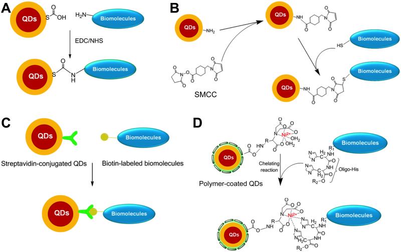
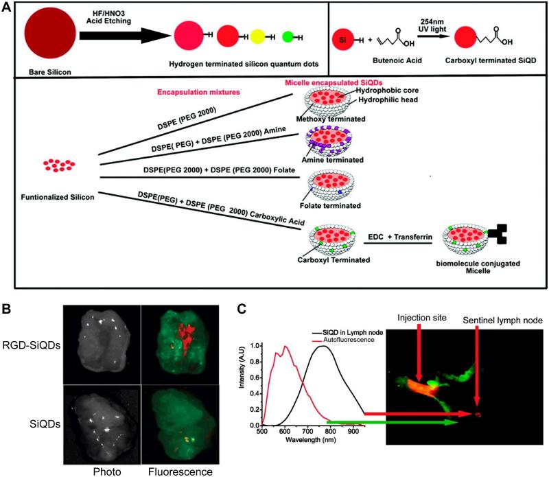
Fluorescent semiconductor quantum dots (QDs) have attracted tremendous attention over the last decade. The superior optical properties of QDs over conventional organic dyes make them attractive labels for a wide variety of biomedical applications, whereas their potential toxicity and instability in biological environment have puzzled scientific researchers. Much research effort has been devoted to surface modification and functionalization of QDs to make them versatile probes for biomedical applications, and significant progress has been made over the last several years. This review article aims to describe the current state-of-the-art of the synthesis, modification, bioconjugation, and applications of QDs for in vivo targeted imaging. In addition, QD-based multifunctional nanoprobes are also summarized.

摘要

在过去的十年中,荧光半导体量子点 (QDs) 引起了极大的关注。QDs 在光学性质上优于传统的有机染料,这使得它们成为各种生物医学应用的有吸引力的标记物,而它们在生物环境中的潜在毒性和不稳定性令科学研究人员感到困惑。人们投入了大量的研究努力来对 QDs 进行表面修饰和功能化,以使它们成为生物医学应用的多功能探针,并且在过去的几年中已经取得了重大进展。本文旨在描述 QDs 的合成、修饰、生物偶联和体内靶向成像应用的最新进展。此外,还总结了基于 QD 的多功能纳米探针。

https://cdn.ncbi.nlm.nih.gov/pmc/blobs/7bd3/3838460/c4cb64b4200b/nihms-523366-f0009.jpg
https://cdn.ncbi.nlm.nih.gov/pmc/blobs/7bd3/3838460/4d5fa19c062c/nihms-523366-f0001.jpg
https://cdn.ncbi.nlm.nih.gov/pmc/blobs/7bd3/3838460/b5b757e6d6bd/nihms-523366-f0002.jpg
https://cdn.ncbi.nlm.nih.gov/pmc/blobs/7bd3/3838460/0a55dcb0e712/nihms-523366-f0003.jpg
https://cdn.ncbi.nlm.nih.gov/pmc/blobs/7bd3/3838460/7f48f75b0376/nihms-523366-f0004.jpg
https://cdn.ncbi.nlm.nih.gov/pmc/blobs/7bd3/3838460/b1781ced52d0/nihms-523366-f0005.jpg
https://cdn.ncbi.nlm.nih.gov/pmc/blobs/7bd3/3838460/68e9c1955464/nihms-523366-f0006.jpg
https://cdn.ncbi.nlm.nih.gov/pmc/blobs/7bd3/3838460/07c41d049aa8/nihms-523366-f0007.jpg
https://cdn.ncbi.nlm.nih.gov/pmc/blobs/7bd3/3838460/bfd8f4c5dbc8/nihms-523366-f0008.jpg
https://cdn.ncbi.nlm.nih.gov/pmc/blobs/7bd3/3838460/c4cb64b4200b/nihms-523366-f0009.jpg