Worth Amy A, Forrest Abigail S, Peri Lauren E, Ward Sean M, Hennig Grant W, Sanders Kenton M
Faculty of Life Sciences, University of Manchester, Manchester, United Kingdom.
Charles Rivers Laboratory, Reno, Nevada, USA.
J Neurogastroenterol Motil. 2015 Mar 30;21(2):200-16. doi: 10.5056/jnm14120.
BACKGROUND/AIMS: Gastric peristalsis begins in the orad corpus and propagates to the pylorus. Directionality of peristalsis depends upon orderly generation and propagation of electrical slow waves and a frequency gradient between proximal and distal pacemakers. We sought to understand how chronotropic agonists affect coupling between corpus and antrum.
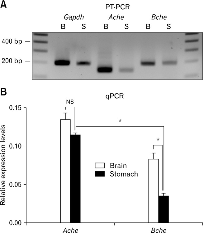
Electrophysiological and imaging techniques were used to investigate regulation of gastric slow wave frequency by muscarinic agonists in mice. We also investigated the expression and role of cholinesterases in regulating slow wave frequency and motor patterns in the stomach.
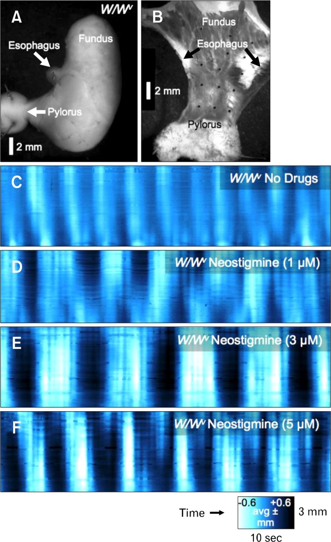
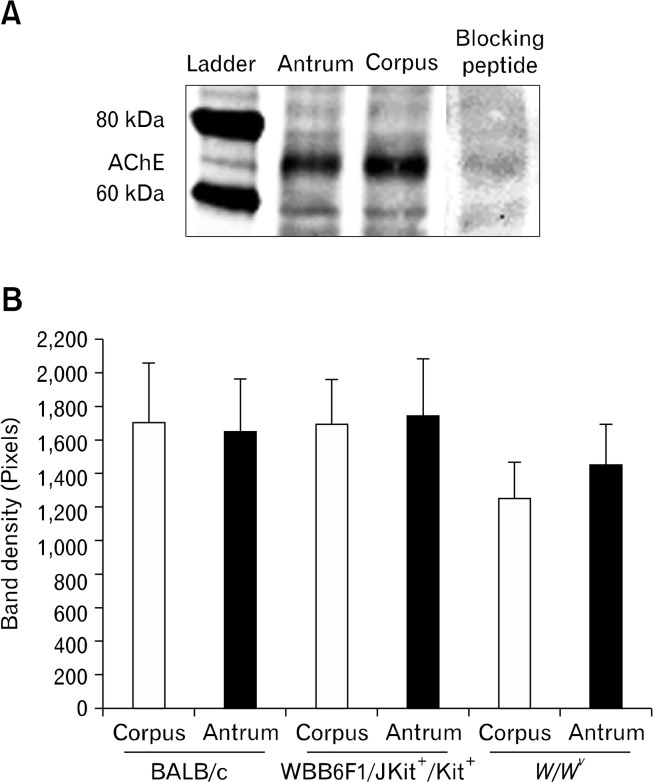
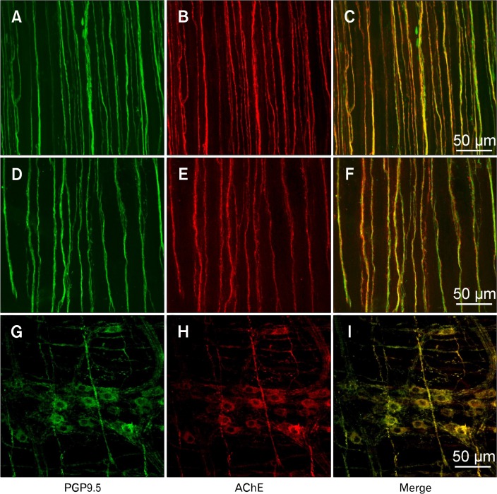
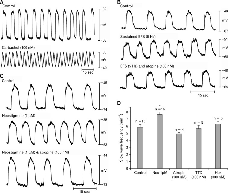
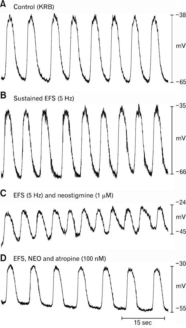
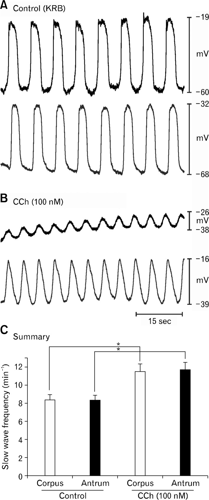
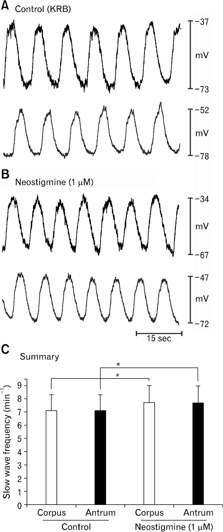
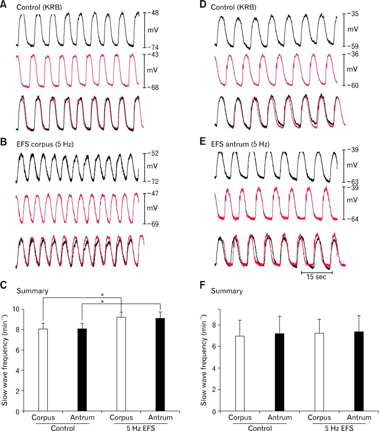
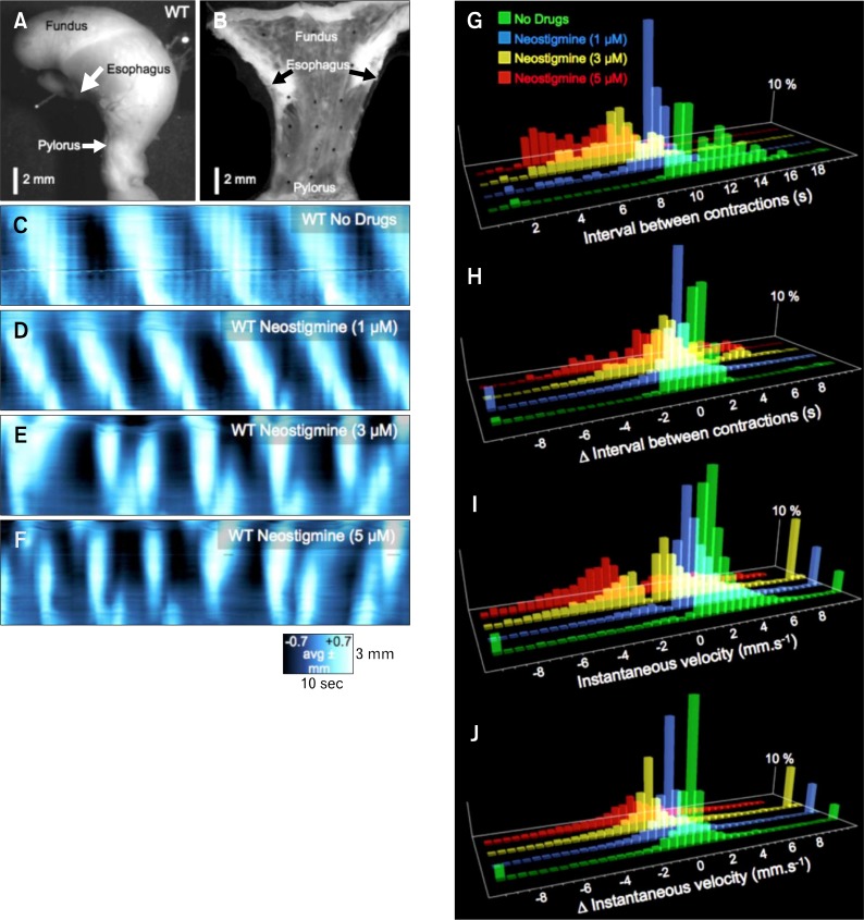
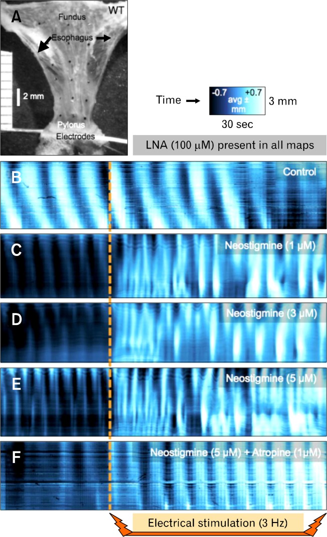
Both acetycholinesterase (Ache) and butyrylcholine esterase (Bche) are expressed in gastric muscles and AChE is localized to var-icose processes of motor neurons. Inhibition of AChE in the absence of stimulation increased slow wave frequency in corpus and throughout muscle strips containing corpus and antrum. CCh caused depolarization and increased slow wave frequency. Stimulation of cholinergic neurons increased slow wave frequency but did not cause depolarization. Neostigmine (1 μM) in-creased slow wave frequency, but uncoupling between corpus and antrum was not detected. Motility mapping of contractile activity in gastric muscles showed similar effects of enteric nerve stimulation on the frequency and propagation of slow waves, but neostigmine (> 1 μM) caused aberrant contractile frequency and propagation and ectopic pacemaking.
Our data show that slow wave uncoupling is difficult to assess with electrical recording from a single or double sites and sug-gest that efficient metabolism of ACh released from motor neurons is an extremely important regulator of slow wave frequency and propagation and gastric motility patterns.
背景/目的:胃蠕动始于胃体近端并向幽门传播。蠕动的方向性取决于电慢波的有序产生和传播以及近端和远端起搏点之间的频率梯度。我们试图了解变时性激动剂如何影响胃体和胃窦之间的耦合。
采用电生理和成像技术研究毒蕈碱激动剂对小鼠胃慢波频率的调节。我们还研究了胆碱酯酶在调节胃慢波频率和运动模式中的表达及作用。
乙酰胆碱酯酶(Ache)和丁酰胆碱酯酶(Bche)均在胃肌中表达,且AChE定位于运动神经元的曲张突起。在无刺激情况下抑制AChE可增加胃体以及包含胃体和胃窦的整个肌条中的慢波频率。氯化乙酰胆碱(CCh)引起去极化并增加慢波频率。刺激胆碱能神经元增加慢波频率,但未引起去极化。新斯的明(1μM)增加慢波频率,但未检测到胃体和胃窦之间的解耦联。胃肌收缩活动的运动图谱显示,肠神经刺激对慢波频率和传播有类似影响,但新斯的明(>1μM)导致异常的收缩频率、传播和异位起搏。
我们的数据表明,通过单部位或双部位电记录很难评估慢波解耦联,并提示运动神经元释放的乙酰胆碱的有效代谢是慢波频率、传播和胃运动模式的极其重要的调节因子。