Losi Gabriele, Marcon Iacopo, Mariotti Letizia, Sessolo Michele, Chiavegato Angela, Carmignoto Giorgio
Neuroscience Institute, National Research Council (CNR) and Department of Biomedical Sciences, University of Padova, via U. Bassi 58/b, 35121 Padova, Italy.
Neuroscience Institute, National Research Council (CNR) and Department of Biomedical Sciences, University of Padova, via U. Bassi 58/b, 35121 Padova, Italy.
J Neurosci Methods. 2016 Feb 15;260:125-31. doi: 10.1016/j.jneumeth.2015.04.001. Epub 2015 Apr 8.
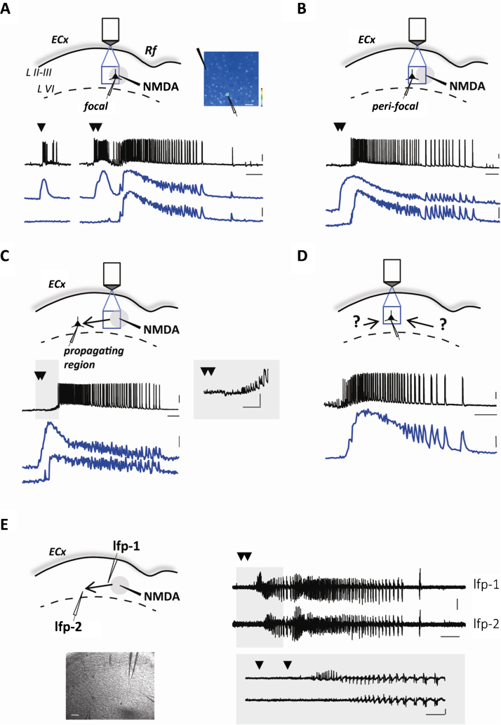
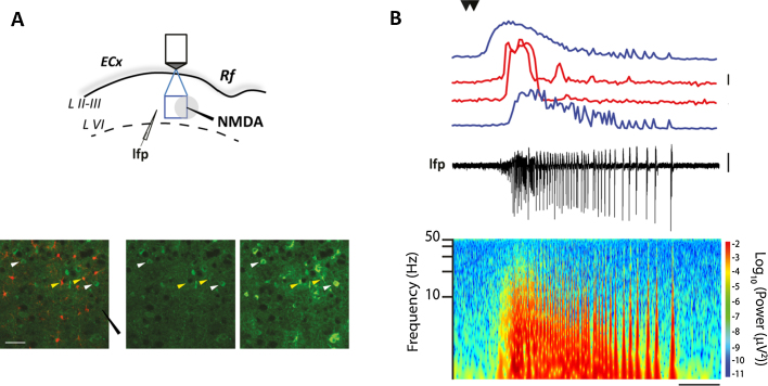
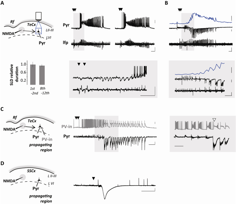
The early cellular events that in a brain network lead to seizure generation and govern seizure propagation are probably based on different cellular mechanisms. Experimental models in which these events can be separately studied would contribute to improve our understanding of epilepsy. We recently described an in vitro model in entorhinal cortex slices from young rats in which focal seizure-like discharges (SLDs) can be induced in spatially defined regions and at predictable times by local NMDA applications performed in the presence of 4-amimopyridine (4-AP) and low extracellular Mg(2+). Through the use of single-dual cell patch-clamp and field potential recordings, and Ca(2+) imaging from large ensembles of neurons, interneurons and astrocytes, we here extend this model to entorhinal and temporal cortex slices of rat and mouse brain, providing evidence that multiple SLDs exhibiting the typical tonic-clonic discharge pattern can be also evoked in these cortical regions by successive NMDA applications. Importantly, the temporal cortex is more accessible to viral vector injections than the entorhinal cortex: this makes it feasible in the former region the selective expression in inhibitory interneurons or principal neurons of genetically encoded Ca(2+) indicators (GECI) or light-gated opsins. In this model, an optogenetic approach allows to activate specific neuronal types at spatially defined locations, i.e., the focus or the propagating region, and at precise time, i.e., before or during SLD. The NMDA/4-AP model can, therefore, represent a valuable tool to gain insights into the role of specific cell populations in seizure generation, propagation and cessation.
在脑网络中导致癫痫发作产生并控制癫痫发作传播的早期细胞事件可能基于不同的细胞机制。能够分别研究这些事件的实验模型将有助于提高我们对癫痫的理解。我们最近描述了一种来自幼鼠内嗅皮层切片的体外模型,在该模型中,通过在4-氨基吡啶(4-AP)和低细胞外镁离子(Mg²⁺)存在的情况下进行局部N-甲基-D-天冬氨酸(NMDA)应用,可以在空间限定区域和可预测的时间诱导局灶性癫痫样放电(SLD)。通过使用单双细胞膜片钳和场电位记录,以及对大量神经元、中间神经元和星形胶质细胞进行钙离子(Ca²⁺)成像,我们在此将该模型扩展到大鼠和小鼠脑的内嗅皮层和颞叶皮层切片,提供证据表明通过连续的NMDA应用,在这些皮质区域也可以诱发表现出典型强直-阵挛放电模式的多个SLD。重要的是,颞叶皮层比内嗅皮层更容易进行病毒载体注射:这使得在前一个区域选择性地在抑制性中间神经元或主要神经元中表达基因编码的钙离子指示剂(GECI)或光门控视蛋白成为可能。在这个模型中,光遗传学方法允许在空间限定位置,即病灶或传播区域,以及精确的时间,即在SLD之前或期间激活特定的神经元类型。因此,NMDA/4-AP模型可以成为一个有价值的工具,以深入了解特定细胞群在癫痫发作产生、传播和终止中的作用。