Kim Jae Heon, Lee Hong Jun, Doo Seung Hwan, Yang Won Jae, Choi Dongho, Kim Jung Hoon, Won Jong Ho, Song Yun Seob
Department of Urology, Soonchunhyang University Seoul Hospital, Soonchunhyang University College of Medicine, Seoul, Korea.
Medical Research Institute, Chung-Ang University College of Medicine, Seoul, Korea.
Korean J Urol. 2015 Apr;56(4):280-7. doi: 10.4111/kju.2015.56.4.280. Epub 2015 Mar 20.
This study was performed to examine the treatment of erectile dysfunction by use of superparamagnetic iron oxide nanoparticles-labeled human mesenchymal stem cells (SPION-MSCs) transplanted into the cavernous nerve injured cavernosa of rats as monitored by molecular magnetic resonance imaging (MRI).
Eight-week-old male Sprague-Dawley rats were divided into three groups of 10 rats each: group 1, sham operation; group 2, cavernous nerve injury; group 3, SPION-MSC treatment after cavernous nerve injury. Immediately after the cavernous nerve injury in group 3, SPION-MSCs were injected into the cavernous nerve injured cavernosa. Serial T2-weighted MRI was done immediately after injection and at 2 and 4 weeks. Erectile response was assessed by cavernous nerve stimulation at 2 and 4 weeks.
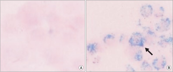
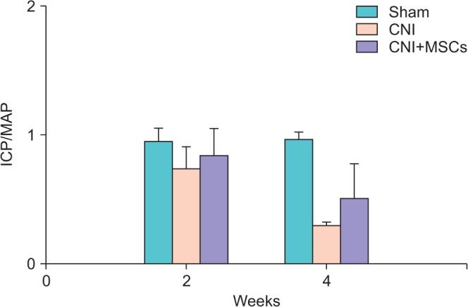
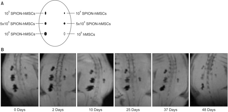
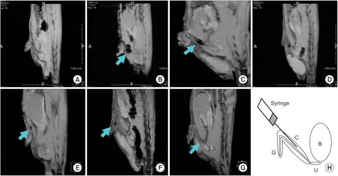
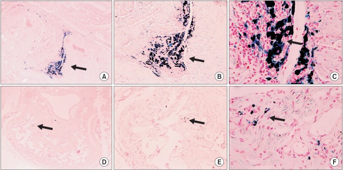
Prussian blue staining of SPION-MSCs revealed abundant uptake of SPION in the cytoplasm. After injection of 1×10(6) SPION-MSCs into the cavernosa of rats, T2-weighted MRI showed a clear hypointense signal induced by the injection. The presence of SPION in the corpora cavernosa was confirmed with Prussian blue staining. At 2 and 4 weeks, rats with cavernous nerve injury had significantly lower erectile function than did rats without cavernous nerve injury (p<0.05). The group transplanted with SPION-MSCs showed higher erectile function than did the group without SPION-MSCs (p<0.05). The presence of SPION-MSCs for up to 4 weeks was confirmed by MRI imaging and Prussian blue staining in the corpus cavernosa.
Transplanted SPION-MSCs existed for up to 4 weeks in the cavernous nerve injured cavernosa of rats. Erectile dysfunction recovered and could be monitored by MRI.
本研究旨在通过分子磁共振成像(MRI)监测,研究超顺磁性氧化铁纳米颗粒标记的人间充质干细胞(SPION-MSCs)移植到大鼠海绵体神经损伤的海绵体中治疗勃起功能障碍的效果。
8周龄雄性Sprague-Dawley大鼠分为三组,每组10只:第1组,假手术;第2组,海绵体神经损伤;第3组,海绵体神经损伤后SPION-MSC治疗。第3组在海绵体神经损伤后立即将SPION-MSCs注入海绵体神经损伤的海绵体中。注射后立即以及在2周和4周时进行系列T2加权MRI检查。在2周和4周时通过海绵体神经刺激评估勃起反应。
SPION-MSCs的普鲁士蓝染色显示细胞质中大量摄取了SPION。向大鼠海绵体注射1×10(6)个SPION-MSCs后,T2加权MRI显示注射诱导出明显的低信号。普鲁士蓝染色证实海绵体中存在SPION。在2周和4周时,海绵体神经损伤的大鼠勃起功能明显低于未损伤海绵体神经的大鼠(p<0.05)。移植SPION-MSCs的组比未移植SPION-MSCs的组勃起功能更高(p<0.05)。MRI成像和普鲁士蓝染色证实海绵体中存在SPION-MSCs长达4周。
移植的SPION-MSCs在大鼠海绵体神经损伤的海绵体中存在长达4周。勃起功能障碍得到恢复,并且可以通过MRI进行监测。