Poché Ross A, Hsu Chih-Wei, McElwee Melissa L, Burns Alan R, Dickinson Mary E
Department of Molecular Physiology and Biophysics, Baylor College of Medicine, Houston, TX, United States; Integrative Molecular and Biomedical Sciences Graduate Program, Baylor College of Medicine, Houston, TX, United States.
Department of Molecular Physiology and Biophysics, Baylor College of Medicine, Houston, TX, United States.
Dev Biol. 2015 Jul 1;403(1):30-42. doi: 10.1016/j.ydbio.2015.03.017. Epub 2015 Apr 24.
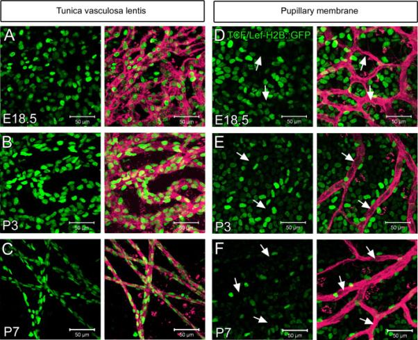
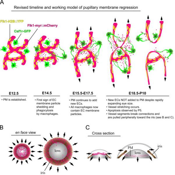
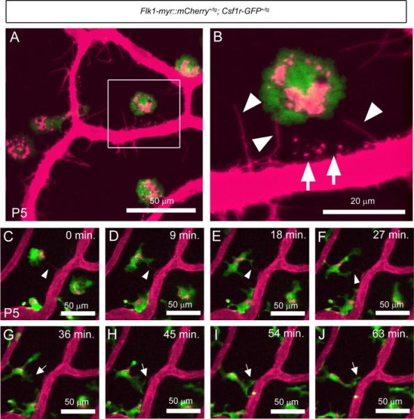
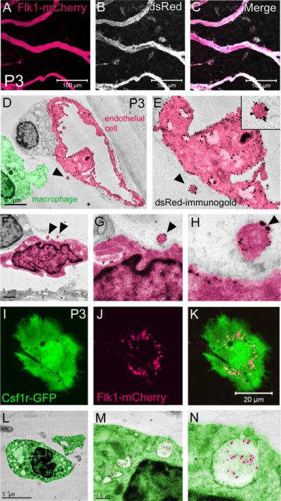
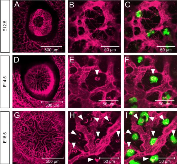
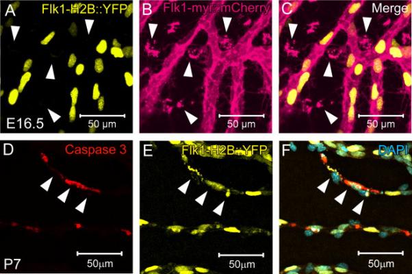
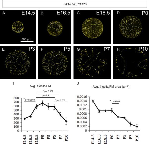
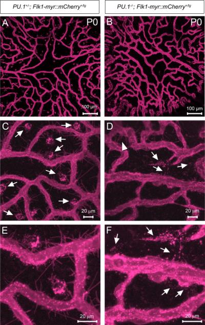
Programmed capillary regression and remodeling are essential developmental processes. However, the cellular and molecular mechanisms that regulate vessel regression are only the beginning to be understood. Here, using in vivo, dynamic, confocal imaging of mouse transgenic reporters as well as static confocal and electron microscopy, we studied the embryonic development and postnatal regression of the transient mouse pupillary membrane (PM) vasculature. This approach allowed us to directly observe the precise temporal sequence of cellular events preceding and during the elimination of the PM from the mouse eye. Imaging of Tcf/Lef-H2B::GFP Wnt-reporter mice uncovered that, unlike the hyaloid vasculature of the posterior eye, a PM endothelial cell (EC) Wnt/β-catenin response is unlikely to be part of the regression mechanism. Live imaging of EC and macrophage dynamics revealed highly active Csf1r-GFP+ macrophages making direct contact with the Flk1-myr::mCherry+ vessel surface and with membrane protrusions or filopodia extending from the ECs. Flk1-myr::mCherry+ EC membrane particles were observed on and around ECs as well as within macrophages. Electron microscopy studies confirmed that they were in phagosomes within macrophages, indicating that the macrophages engulfed the membrane particles. Interestingly, EC plasma membrane uptake by PM macrophages did not correlate with apoptosis and was found shortly after vessel formation at mid-gestation stages in the embryo; long before vessel regression begins during postnatal development. Additionally, genetic ablation of macrophages showed that EC membrane particles were still shed in the absence of macrophages suggesting that macrophages do not induce the formation or release of EC microparticles. These studies have uncovered a novel event during programmed capillary regression in which resident macrophages scavenge endothelial cell microparticles released from the PM vessels. This finding suggests that there may be an initial disruption in vessel homeostasis embryonically as the PM forms that may underlie its ultimate regression postnatally.
程序性毛细血管消退和重塑是重要的发育过程。然而,调节血管消退的细胞和分子机制才刚刚开始被了解。在这里,我们利用小鼠转基因报告基因的体内动态共聚焦成像以及静态共聚焦和电子显微镜,研究了小鼠瞬态瞳孔膜(PM)脉管系统的胚胎发育和出生后消退。这种方法使我们能够直接观察在小鼠眼中PM消除之前和期间细胞事件的精确时间顺序。对Tcf/Lef-H2B::GFP Wnt报告基因小鼠的成像发现,与后眼的玻璃体脉管系统不同,PM内皮细胞(EC)的Wnt/β-连环蛋白反应不太可能是消退机制的一部分。对EC和巨噬细胞动态的实时成像显示,高度活跃的Csf1r-GFP+巨噬细胞与Flk1-myr::mCherry+血管表面直接接触,并与从EC延伸的膜突起或丝状伪足接触。在EC上、周围以及巨噬细胞内观察到Flk1-myr::mCherry+ EC膜颗粒。电子显微镜研究证实它们存在于巨噬细胞内的吞噬体中,表明巨噬细胞吞噬了膜颗粒。有趣的是,PM巨噬细胞对EC质膜的摄取与细胞凋亡无关,并且在胚胎中期血管形成后不久就被发现;远早于出生后发育期间血管消退开始之前。此外,巨噬细胞的基因消融表明,在没有巨噬细胞的情况下,EC膜颗粒仍然会脱落,这表明巨噬细胞不会诱导EC微粒的形成或释放。这些研究揭示了程序性毛细血管消退过程中的一个新事件,即驻留巨噬细胞清除从PM血管释放的内皮细胞微粒。这一发现表明,在胚胎期PM形成时,血管稳态可能会出现初始破坏,这可能是其出生后最终消退的基础。