• 文献检索
  • 文档翻译
  • 深度研究
  • 学术资讯
  • Suppr Zotero 插件Zotero 插件
  • 邀请有礼
  • 套餐&价格
  • 历史记录
应用&插件
Suppr Zotero 插件Zotero 插件浏览器插件Mac 客户端Windows 客户端微信小程序
定价
高级版会员购买积分包购买API积分包
服务
文献检索文档翻译深度研究API 文档MCP 服务
关于我们
关于 Suppr公司介绍联系我们用户协议隐私条款
关注我们

Suppr 超能文献

核心技术专利:CN118964589B侵权必究
粤ICP备2023148730 号-1Suppr @ 2026

文献检索

告别复杂PubMed语法,用中文像聊天一样搜索,搜遍4000万医学文献。AI智能推荐,让科研检索更轻松。

立即免费搜索

文件翻译

保留排版,准确专业,支持PDF/Word/PPT等文件格式,支持 12+语言互译。

免费翻译文档

深度研究

AI帮你快速写综述,25分钟生成高质量综述,智能提取关键信息,辅助科研写作。

立即免费体验

光学相干断层扫描中高反射点的病因,不包括糖尿病性黄斑水肿和视网膜静脉阻塞§

The causes of hyperreflective dots in optical coherence tomography excluding diabetic macular edema and retinal venous occlusion§.

作者信息

Turgut Burak, Yildirim Hakan

机构信息

Fırat University, School of Medicine, Department of Ophthalmology, Elazığ, Turkey.

出版信息

Open Ophthalmol J. 2015 Mar 31;9:36-40. doi: 10.2174/1874364101509010036. eCollection 2015.

DOI:10.2174/1874364101509010036
PMID:25926902
原文链接:https://pmc.ncbi.nlm.nih.gov/articles/PMC4407005/
Abstract

PURPOSE

To investigate the causes of hyperreflective dots (HRDs) in spectral domain optical coherence tomography (OCT) excluding diabetic macular edema (DME) and RVO (retinal vein occlusion).

PATIENTS AND METHODS

The medical records of 56 patients with HRDs documented by OCT were reviewed retrospectively. The patients with DME and RVO were excluded from the study in order to prevent misdiagnosing hard exudates or HRDs. The causes, unilaterality or bilaterality of HRD and demographic properties of the patients with HRD were evaluated.

RESULTS

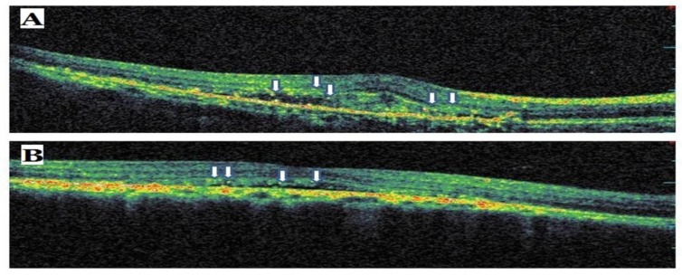
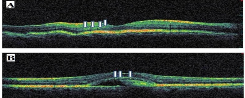
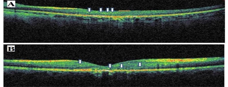
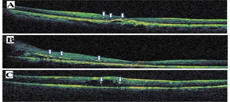
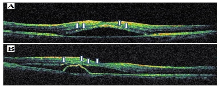
Sixty four eyes of 56 patients having HRDs were included in this study. Of the patients with HRD, 17 (30.36%) were women and 39 (69.64%) were men. The ages of patients were between 13 to 84 years (median 60.18 years). The causes of HRD were as follows: papilledema in 4 eyes (6.25%), active neovascular age related macular degeneration (AMD) in 33 eyes (51.56%), familial dominant drusen in 2 eyes (3.13%), central serous chorioretinopathy in 19 eyes (29.69%) and commotio retina in 2 eyes (3.13%), choroidal folds in one eye (1.56%), branch retinal artery occlusion in one eye (1.56%), central retinal artery occlusion in one patient (1.56%) and Best vitelliform macular dystrophy in one eye (1.56%). The most common cause of HRD was AMD. The causes of HRDs in both eyes were AMD and papilledema.

CONCLUSION

The most common causes of HRDs excluding DME and RVO seem as active exudative AMD. The presence of HRDs in retinal diseases might affect the decisions and the results of the treatment and the prognosis of diseases.

摘要

目的

探讨光谱域光学相干断层扫描(OCT)中高反射点(HRDs)的病因,排除糖尿病性黄斑水肿(DME)和视网膜静脉阻塞(RVO)。

患者与方法

回顾性分析56例经OCT记录有HRDs患者的病历。为防止误诊硬性渗出或HRDs,将患有DME和RVO的患者排除在研究之外。评估HRD的病因、单眼或双眼情况以及HRD患者的人口统计学特征。

结果

本研究纳入了56例有HRDs的患者的64只眼。在有HRD的患者中,女性17例(30.36%),男性39例(69.64%)。患者年龄在13至84岁之间(中位数60.18岁)。HRD的病因如下:视乳头水肿4只眼(6.25%),活动性新生血管性年龄相关性黄斑变性(AMD)33只眼(51.56%),家族性显性玻璃膜疣2只眼(3.13%)中央性浆液性脉络膜视网膜病变19只眼(29.69%),视网膜震荡2只眼(3.13%),脉络膜皱褶1只眼(1.56%),视网膜分支动脉阻塞1只眼(1.56%),视网膜中央动脉阻塞1例(1.56%),Best卵黄状黄斑营养不良1只眼(1.56%)。HRD最常见的病因是AMD。双眼HRD的病因是AMD和视乳头水肿。

结论

排除DME和RVO后,HRDs最常见的病因似乎是活动性渗出性AMD。视网膜疾病中HRDs的存在可能会影响治疗决策、治疗结果和疾病预后。

https://cdn.ncbi.nlm.nih.gov/pmc/blobs/5c17/4407005/f5c480c40487/TOOPHTJ-9-36_F5.jpg
https://cdn.ncbi.nlm.nih.gov/pmc/blobs/5c17/4407005/a9d101d58023/TOOPHTJ-9-36_F1.jpg
https://cdn.ncbi.nlm.nih.gov/pmc/blobs/5c17/4407005/17c0db10aa19/TOOPHTJ-9-36_F2.jpg
https://cdn.ncbi.nlm.nih.gov/pmc/blobs/5c17/4407005/cd9cbead47b4/TOOPHTJ-9-36_F3.jpg
https://cdn.ncbi.nlm.nih.gov/pmc/blobs/5c17/4407005/13f15d7e08fb/TOOPHTJ-9-36_F4.jpg
https://cdn.ncbi.nlm.nih.gov/pmc/blobs/5c17/4407005/f5c480c40487/TOOPHTJ-9-36_F5.jpg

相似文献

1
The causes of hyperreflective dots in optical coherence tomography excluding diabetic macular edema and retinal venous occlusion§.光学相干断层扫描中高反射点的病因,不包括糖尿病性黄斑水肿和视网膜静脉阻塞§
Open Ophthalmol J. 2015 Mar 31;9:36-40. doi: 10.2174/1874364101509010036. eCollection 2015.
2
Association Between Hyperreflective Dots on Spectral-Domain Optical Coherence Tomography in Macular Edema and Response to Treatment.黄斑水肿患者光谱域光学相干断层扫描中高反射点与治疗反应之间的关联
Invest Ophthalmol Vis Sci. 2017 Nov 1;58(13):5958-5967. doi: 10.1167/iovs.17-22725.
3
Predictive capacity of baseline hyperreflective dots on the intravitreal dexamethasone implant (Ozurdex®) outcomes in diabetic macular edema: a multicenter study.基线高反射点对玻璃体内曲安奈德植入物(Ozurdex®)治疗糖尿病性黄斑水肿结局的预测能力:一项多中心研究。
Graefes Arch Clin Exp Ophthalmol. 2019 Nov;257(11):2381-2390. doi: 10.1007/s00417-019-04446-4. Epub 2019 Aug 26.
4
Hyperreflective Dots on OCT as a Predictor of Treatment Outcome in Diabetic Macular Edema: A Systematic Review.OCT 上的高反射点预测糖尿病性黄斑水肿的治疗效果:系统评价。
Ophthalmol Retina. 2022 Sep;6(9):814-827. doi: 10.1016/j.oret.2022.03.020. Epub 2022 Mar 30.
5
Optical coherence tomographic evaluation of foveal hard exudates in patients with diabetic maculopathy accompanying macular detachment.光学相干断层扫描评价伴有黄斑脱离的糖尿病性黄斑病变患者的中心凹硬性渗出物。
Ophthalmology. 2010 Oct;117(10):1996-2002. doi: 10.1016/j.ophtha.2010.06.019. Epub 2010 Aug 17.
6
Comparing retinal thickness measurements using automated fast macular thickness map versus six-radial line scans with manual measurements.比较使用自动快速黄斑厚度图与六径向线扫描及手动测量的视网膜厚度测量结果。
Ophthalmology. 2009 May;116(5):964-70. doi: 10.1016/j.ophtha.2008.12.033.
7
Algorithm for Detection and Quantification of Hyperreflective Dots on Optical Coherence Tomography in Diabetic Macular Edema.糖尿病性黄斑水肿光学相干断层扫描中高反射点检测与定量算法
Front Med (Lausanne). 2021 Aug 18;8:688986. doi: 10.3389/fmed.2021.688986. eCollection 2021.
8
Evaluation of choroidal hyperreflective dots in acute and chronic central serous chorioretinopathy.评价急性和慢性中心性浆液性脉络膜视网膜病变的脉络膜高反射点。
Indian J Ophthalmol. 2019 Nov;67(11):1850-1854. doi: 10.4103/ijo.IJO_2030_18.
9
Characteristics of optical coherence tomographic hyperreflective foci in retinal vein occlusion.视网膜静脉阻塞的光相干断层扫描高反射病灶的特征。
Retina. 2012 Jan;32(1):77-85. doi: 10.1097/IAE.0b013e318217ffc7.
10
Pseudoflow with OCT Angiography in Eyes with Hard Exudates and Macular Drusen.伴有硬性渗出和黄斑玻璃膜疣的眼中光学相干断层扫描血管造影的伪血流现象
Transl Vis Sci Technol. 2019 Jun 27;8(3):50. doi: 10.1167/tvst.8.3.50. eCollection 2019 May.

引用本文的文献

1
Hyperreflective Retinal Foci (HRF): Definition and Role of an Invaluable OCT Sign.高反射性视网膜病灶(HRF):一种重要的光学相干断层扫描(OCT)征象的定义及作用
J Clin Med. 2025 Apr 27;14(9):3021. doi: 10.3390/jcm14093021.
2
Intravitreal Administration of Avacincaptad Pegol in a Nonhuman Primate Model of Dry Age-Related Macular Degeneration.在干性年龄相关性黄斑变性非人灵长类动物模型中玻璃体内注射阿伐西普聚乙二醇化产物
Pharmacol Res Perspect. 2025 Feb;13(1):e70052. doi: 10.1002/prp2.70052.
3
Comparison between spectral-domain optical coherence tomography-based and partial coherence interferometry-based biometry.

本文引用的文献

1
Hyperreflective intraretinal spots in diabetics without and with nonproliferative diabetic retinopathy: an in vivo study using spectral domain OCT.糖尿病患者无和伴非增生性糖尿病视网膜病变的视网膜内高反射性斑点:应用谱域 OCT 的活体研究。
J Diabetes Res. 2013;2013:491835. doi: 10.1155/2013/491835. Epub 2013 Dec 9.
2
Optical coherence tomography--current and future applications.光学相干断层扫描——当前和未来的应用。
Curr Opin Ophthalmol. 2013 May;24(3):213-21. doi: 10.1097/ICU.0b013e32835f8bf8.
3
Hyperreflective dots: a new spectral-domain optical coherence tomography entity for follow-up and prognosis in exudative age-related macular degeneration.
谱域光学相干断层扫描生物测量与部分相干干涉测量生物测量的比较。
Sci Rep. 2024 Nov 18;14(1):28440. doi: 10.1038/s41598-024-79679-3.
4
Hyperreflective Dots in Central Fovea Visualized by a Novel Application of Visible-Light Optical Coherence Tomography.通过可见光光学相干断层扫描新技术观察到的中央凹高反射点
Case Rep Ophthalmol Med. 2024 Jul 9;2024:5823455. doi: 10.1155/2024/5823455. eCollection 2024.
5
SARS-CoV-2 neurovascular invasion supported by Mendelian randomization.基于孟德尔随机化的 SARS-CoV-2 神经血管侵犯。
J Transl Med. 2024 Jan 24;22(1):101. doi: 10.1186/s12967-024-04907-3.
6
Hyper-reflective dots in optical coherence tomography imaging and inflammation markers in diabetic retinopathy.光学相干断层扫描成像中的高反射点与糖尿病视网膜病变中的炎症标志物。
J Anat. 2023 Oct;243(4):697-705. doi: 10.1111/joa.13889. Epub 2023 May 24.
7
Aggravation of retinal hard exudates after intravitreal anti-vascular endothelial growth factor therapy for cystoid macular edema and the risk factors: a retrospective study.眼内抗血管内皮生长因子治疗后视网膜硬性渗出加重与黄斑囊样水肿的关系及危险因素:一项回顾性研究。
BMC Ophthalmol. 2022 Feb 23;22(1):92. doi: 10.1186/s12886-022-02315-z.
8
Longitudinal Evaluation of Hyper-Reflective Foci in the Retina Following Subretinal Delivery of Adeno-Associated Virus in Non-Human Primates.视网膜下注射腺相关病毒后猴眼内高反射病灶的纵向评估。
Transl Vis Sci Technol. 2021 May 3;10(6):15. doi: 10.1167/tvst.10.6.15.
9
The Correlation between Hemoglobin A1c (HbA1c) and Hyperreflective Dots (HRD) in Diabetic Patients.糖尿病患者的血红蛋白 A1c(HbA1c)与高反射点(HRD)之间的相关性。
Int J Environ Res Public Health. 2020 May 1;17(9):3154. doi: 10.3390/ijerph17093154.
10
Tracing the retina to analyze the integrity and phagocytic capacity of the retinal pigment epithelium.追踪视网膜以分析视网膜色素上皮的完整性和吞噬能力。
Sci Rep. 2020 Apr 29;10(1):7273. doi: 10.1038/s41598-020-64131-z.
高反射点:一种新的用于渗出性年龄相关性黄斑变性随访和预后的谱域光相干断层扫描实体。
Ophthalmologica. 2013;229(1):32-7. doi: 10.1159/000342159. Epub 2012 Sep 19.
4
Behavior of SD-OCT-detected hyperreflective foci in the retina of anti-VEGF-treated patients with diabetic macular edema.抗 VEGF 治疗糖尿病性黄斑水肿患者的视网膜中 SD-OCT 检测到的高反射焦点的行为。
Invest Ophthalmol Vis Sci. 2012 Aug 24;53(9):5814-8. doi: 10.1167/iovs.12-9950.
5
Relationship between fluorescein pooling and optical coherence tomographic reflectivity of cystoid spaces in diabetic macular edema.糖尿病性黄斑水肿中囊样间隙的荧光素蓄积与光相干断层扫描反射率的关系。
Ophthalmology. 2012 May;119(5):1047-55. doi: 10.1016/j.ophtha.2011.10.030. Epub 2012 Feb 11.
6
Association between hyperreflective foci in the outer retina, status of photoreceptor layer, and visual acuity in diabetic macular edema.外视网膜高反射病灶与光感受器层状态及糖尿病性黄斑水肿视力的关系。
Am J Ophthalmol. 2012 Apr;153(4):710-7, 717.e1. doi: 10.1016/j.ajo.2011.08.041. Epub 2011 Dec 3.
7
Characteristics of optical coherence tomographic hyperreflective foci in retinal vein occlusion.视网膜静脉阻塞的光相干断层扫描高反射病灶的特征。
Retina. 2012 Jan;32(1):77-85. doi: 10.1097/IAE.0b013e318217ffc7.
8
Microglia/macrophages migrate through retinal epithelium barrier by a transcellular route in diabetic retinopathy: role of PKCζ in the Goto Kakizaki rat model.在糖尿病性视网膜病变中,小胶质细胞/巨噬细胞通过细胞间途径迁移穿过视网膜上皮屏障:PKCζ 在 Goto Kakizaki 大鼠模型中的作用。
Am J Pathol. 2011 Aug;179(2):942-53. doi: 10.1016/j.ajpath.2011.04.018. Epub 2011 Jun 2.
9
Management of retinal vein occlusion--consensus document.视网膜静脉阻塞的管理——共识文件。
Ophthalmologica. 2011;226(1):4-28. doi: 10.1159/000327391. Epub 2011 May 17.
10
Optical coherence tomography: history, current status, and laboratory work.光学相干断层扫描:历史、现状和实验室工作。
Invest Ophthalmol Vis Sci. 2011 Apr 14;52(5):2425-36. doi: 10.1167/iovs.10-6312. Print 2011 Apr.