Department of Anatomy and Cell Biology, School of Dental Medicine, University of Pennsylvania, Philadelphia, PA USA ; Pennsylvania Muscle Institute, University of Pennsylvania, Philadelphia, PA USA.
Skelet Muscle. 2014 Nov 27;4:21. doi: 10.1186/2044-5040-4-21. eCollection 2014.
Histological assessment of skeletal muscle tissue is commonly applied to many areas of skeletal muscle physiological research. Histological parameters including fiber distribution, fiber type, centrally nucleated fibers, and capillary density are all frequently quantified measures of skeletal muscle. These parameters reflect functional properties of muscle and undergo adaptation in many muscle diseases and injuries. While standard operating procedures have been developed to guide analysis of many of these parameters, the software to freely, efficiently, and consistently analyze them is not readily available. In order to provide this service to the muscle research community we developed an open source MATLAB script to analyze immunofluorescent muscle sections incorporating user controls for muscle histological analysis.
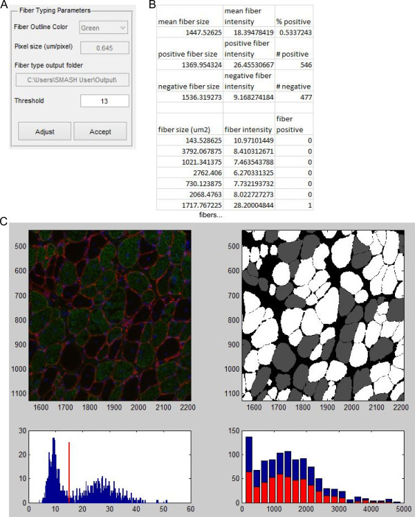
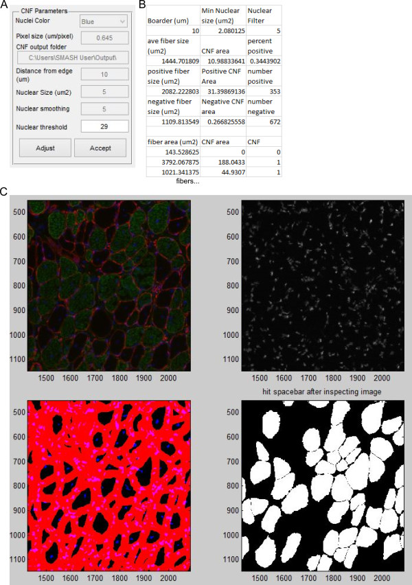
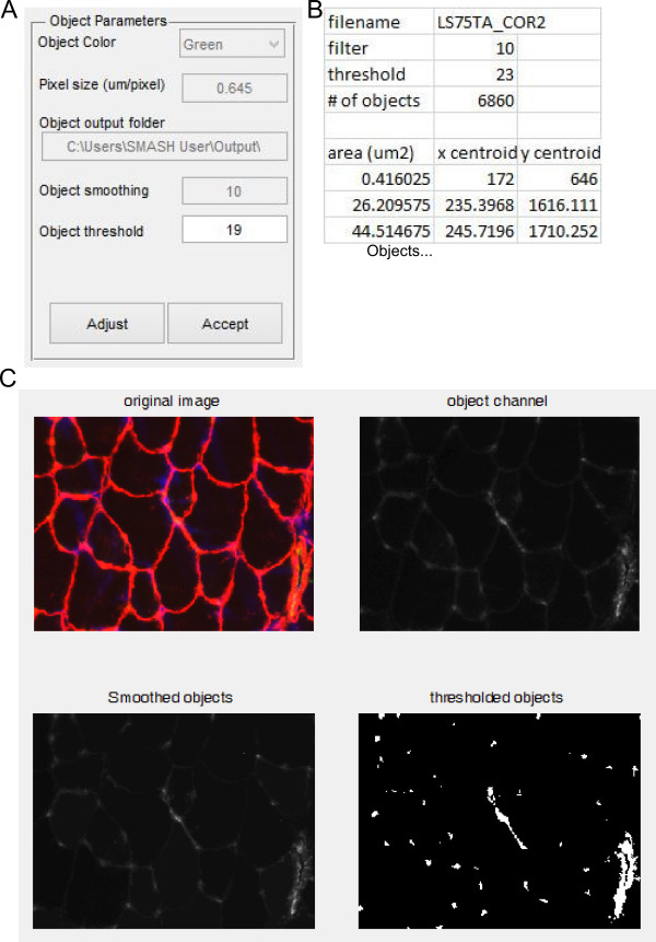
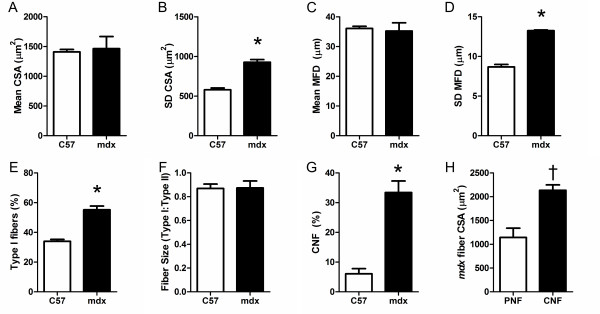
The software consists of multiple functions designed to provide tools for the analysis selected. Initial segmentation and fiber filter functions segment the image and remove non-fiber elements based on user-defined parameters to create a fiber mask. Establishing parameters set by the user, the software outputs data on fiber size and type, centrally nucleated fibers, and other structures. These functions were evaluated on stained soleus muscle sections from 1-year-old wild-type and mdx mice, a model of Duchenne muscular dystrophy. In accordance with previously published data, fiber size was not different between groups, but mdx muscles had much higher fiber size variability. The mdx muscle had a significantly greater proportion of type I fibers, but type I fibers did not change in size relative to type II fibers. Centrally nucleated fibers were highly prevalent in mdx muscle and were significantly larger than peripherally nucleated fibers.
The MATLAB code described and provided along with this manuscript is designed for image processing of skeletal muscle immunofluorescent histological sections. The program allows for semi-automated fiber detection along with user correction. The output of the code provides data in accordance with established standards of practice. The results of the program have been validated using a small set of wild-type and mdx muscle sections. This program is the first freely available and open source image processing program designed to automate analysis of skeletal muscle histological sections.
骨骼肌组织的组织学评估广泛应用于骨骼肌生理学研究的许多领域。包括纤维分布、纤维类型、中央核纤维和毛细血管密度在内的组织学参数都是骨骼肌的常用量化指标。这些参数反映了肌肉的功能特性,并在许多肌肉疾病和损伤中发生适应性变化。虽然已经制定了标准操作规程来指导许多这些参数的分析,但能够自由、高效和一致地分析它们的软件并不容易获得。为了向肌肉研究界提供这项服务,我们开发了一个开源 MATLAB 脚本,用于分析包含用户控制的免疫荧光骨骼肌切片,以进行肌肉组织学分析。
该软件由多个功能组成,旨在为选定的分析提供工具。初始分割和纤维过滤功能根据用户定义的参数分割图像并去除非纤维元素,以创建纤维掩模。该软件根据用户设置的参数输出纤维大小和类型、中央核纤维以及其他结构的数据。这些功能在来自 1 岁野生型和 mdx 小鼠(杜氏肌营养不良症的模型)的染色比目鱼肌切片上进行了评估。与之前发表的数据一致,纤维大小在两组之间没有差异,但 mdx 肌肉的纤维大小变异性要大得多。mdx 肌肉的 I 型纤维比例明显更高,但 I 型纤维相对于 II 型纤维的大小没有变化。中央核纤维在 mdx 肌肉中非常普遍,并且比周围核纤维明显更大。
本文档中描述并提供的 MATLAB 代码旨在处理骨骼肌免疫荧光组织学切片的图像处理。该程序允许进行半自动纤维检测以及用户校正。代码的输出提供了符合既定实践标准的数据。该程序的结果已使用一小部分野生型和 mdx 肌肉切片进行了验证。该程序是第一个专门设计用于自动化分析骨骼肌组织学切片的免费和开源图像处理程序。