Grafft Carrie A, Fervenza Fernando C, Semret Merfake H, Orloff Sheldon, Sethi Sanjeev
Division of Nephrology and Hypertension, Department of Medicine, Mayo Clinic, Rochester, MN.
Kaiser Medical Center, Oakland, CA.
NDT Plus. 2009 Dec;2(6):448-51. doi: 10.1093/ndtplus/sfp101. Epub 2009 Aug 10.
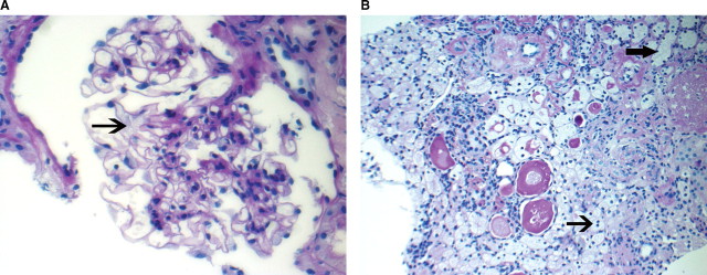
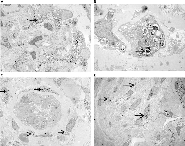
We describe the renal biopsy findings in a 14-year-old girl with Neimann-Pick disease. The renal biopsy showed chronic changes involving all components of the parenchyma, including focal global glomerulosclerosis, tubular atrophy, interstitial fibrosis and vascular sclerosis. On light microscopy, significant findings included foamy podocytes, vacuolated tubular epithelial cells and collections of foam cells in the interstitium. Electron microscopy was confirmatory which showed myelin-like inclusions in podocytes, endothelial cells, tubular epithelial cells and small nerves. The findings are similar to Fabry's disease, except that small nerve involvement appears to be unique to Neimann Pick disease.
我们描述了一名患有尼曼-匹克病的14岁女孩的肾活检结果。肾活检显示实质所有成分均有慢性改变,包括局灶性全肾小球硬化、肾小管萎缩、间质纤维化和血管硬化。在光学显微镜下,显著发现包括泡沫状足细胞、空泡化肾小管上皮细胞以及间质中的泡沫细胞聚集。电子显微镜检查证实了这一结果,显示足细胞、内皮细胞、肾小管上皮细胞和小神经中有髓鞘样包涵体。这些发现与法布里病相似,只是小神经受累似乎是尼曼-匹克病所特有的。