• 文献检索
  • 文档翻译
  • 深度研究
  • 学术资讯
  • Suppr Zotero 插件Zotero 插件
  • 邀请有礼
  • 套餐&价格
  • 历史记录
应用&插件
Suppr Zotero 插件Zotero 插件浏览器插件Mac 客户端Windows 客户端微信小程序
定价
高级版会员购买积分包购买API积分包
服务
文献检索文档翻译深度研究API 文档MCP 服务
关于我们
关于 Suppr公司介绍联系我们用户协议隐私条款
关注我们

Suppr 超能文献

核心技术专利:CN118964589B侵权必究
粤ICP备2023148730 号-1Suppr @ 2026

文献检索

告别复杂PubMed语法,用中文像聊天一样搜索,搜遍4000万医学文献。AI智能推荐,让科研检索更轻松。

立即免费搜索

文件翻译

保留排版,准确专业,支持PDF/Word/PPT等文件格式,支持 12+语言互译。

免费翻译文档

深度研究

AI帮你快速写综述,25分钟生成高质量综述,智能提取关键信息,辅助科研写作。

立即免费体验

Post-treatment imaging of high-grade gliomas.

作者信息

Sanghvi Darshana

机构信息

Department of Radiology, Kokilaben Dhirubhai Ambani Hospital, Mumbai, Maharashtra, India.

出版信息

Indian J Radiol Imaging. 2015 Apr-Jun;25(2):102-8. doi: 10.4103/0971-3026.155829.

DOI:10.4103/0971-3026.155829
PMID:25969633
原文链接:https://pmc.ncbi.nlm.nih.gov/articles/PMC4419419/
Abstract

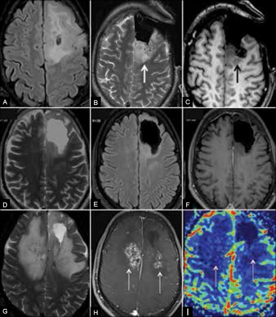
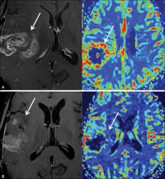
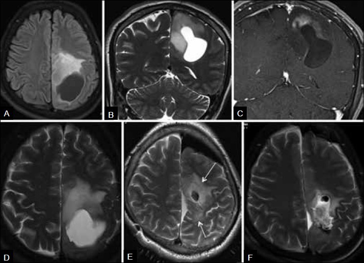
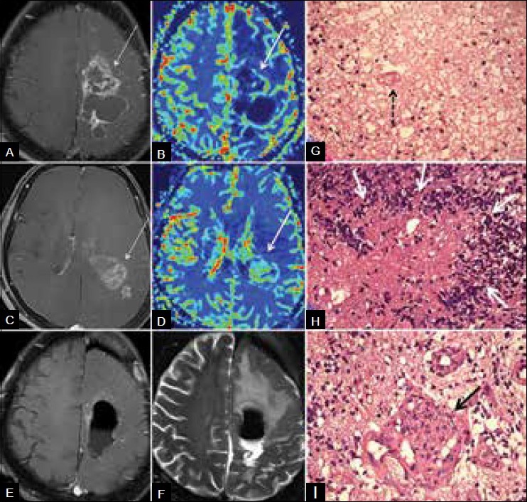
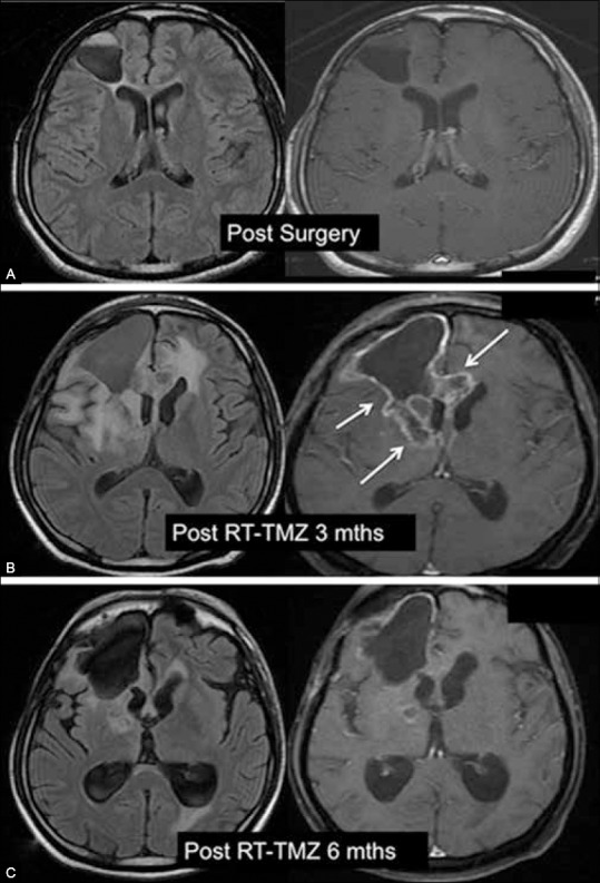
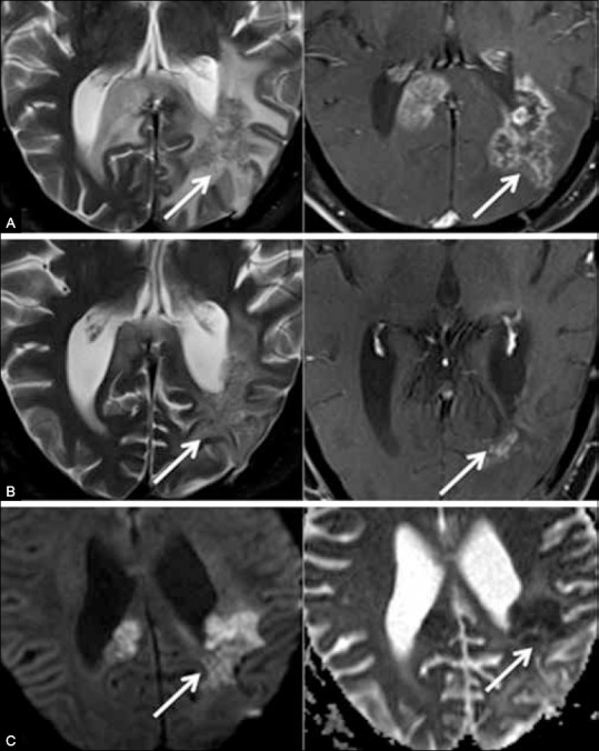
Current standard of care for treatment of newly diagnosed high grade gliomas is surgery followed by concomitant radiotherapy (RT) and chemotherapy (CT) with temozolomide (TMZ). Recently, bevacizumab, an anti - angiogenic agent has also been approved for treatment of recurrent gliomas. Baseline imaging after excision is optimally obtained in the first 24 hours. When baseline postoperative imaging is delayed beyond 24 hours, subacute hemorrhage, subacute ischemia and inflammation at the resection margins render differentiation from residual tumor challenging. Radiation necrosis is a well recognized entity and is differentiated from recurrence based on morphology on structural imaging, presence of lipid - lactate complexes with lack of choline on spectroscopy and low normalized cerebral blood volume (CBV) ratios at perfusion imaging. Novel chemotherapies have lead to the occurrence of interesting but sometimes confusing post treatment imaging appearances including the phenomena of 'pseudoprogression' and 'pseudoresponse'. Pseudoprogression refers to transient, self resolving focal enhancement mediated by TMZ-induced increased vascular permeability and local inflammatory response. Pathologically, these lesions do not have viable tumor. The lesions stabilize or regress without further treatment and are usually clinically asymptomatic. Pseudoresponse refers to rapid regression of enhancement, perfusion, mass effect and midline shift caused by the anti - angiogenic effect of bevacizumab. It is termed pseudoresponse since biological tumor persists as non-enhancing altered signal. It is important for radiologists to be aware of these entities seen on post treatment imaging of gliomas, as misinterpretation may lead to inappropriate management decisions and prognostication.

摘要
https://cdn.ncbi.nlm.nih.gov/pmc/blobs/2baf/4419419/d4ab05218575/IJRI-25-102-g007.jpg
https://cdn.ncbi.nlm.nih.gov/pmc/blobs/2baf/4419419/2318d3650ce8/IJRI-25-102-g001.jpg
https://cdn.ncbi.nlm.nih.gov/pmc/blobs/2baf/4419419/a71dee51a3da/IJRI-25-102-g003.jpg
https://cdn.ncbi.nlm.nih.gov/pmc/blobs/2baf/4419419/3f85ff391ae0/IJRI-25-102-g004.jpg
https://cdn.ncbi.nlm.nih.gov/pmc/blobs/2baf/4419419/bc9881705993/IJRI-25-102-g005.jpg
https://cdn.ncbi.nlm.nih.gov/pmc/blobs/2baf/4419419/70f213d1bec8/IJRI-25-102-g006.jpg
https://cdn.ncbi.nlm.nih.gov/pmc/blobs/2baf/4419419/d4ab05218575/IJRI-25-102-g007.jpg

相似文献

1
Post-treatment imaging of high-grade gliomas.
Indian J Radiol Imaging. 2015 Apr-Jun;25(2):102-8. doi: 10.4103/0971-3026.155829.
2
Radiation Necrosis, Pseudoprogression, Pseudoresponse, and Tumor Recurrence: Imaging Challenges for the Evaluation of Treated Gliomas.放射性坏死、假性进展、假性缓解和肿瘤复发:治疗后脑胶质瘤评估的影像学挑战。
Contrast Media Mol Imaging. 2018 Dec 2;2018:6828396. doi: 10.1155/2018/6828396. eCollection 2018.
3
Pseudoprogression after radiotherapy with concurrent temozolomide for high-grade glioma: clinical observations and working recommendations.同步替莫唑胺放疗后高级别胶质瘤的假性进展:临床观察与工作建议
Surg Neurol. 2009 Oct;72(4):423-8. doi: 10.1016/j.surneu.2008.09.023. Epub 2009 Jan 15.
4
Pseudoprogression: relevance with respect to treatment of high-grade gliomas.假性进展:与高级别胶质瘤治疗的相关性。
Curr Treat Options Oncol. 2011 Sep;12(3):240-52. doi: 10.1007/s11864-011-0157-1.
5
Pseudoprogression and pseudoresponse in the management of high-grade glioma : optimal decision timing according to the response assessment of the neuro-oncology working group.高级别胶质瘤治疗中的假性进展和假性反应:根据神经肿瘤学工作组的反应评估确定最佳决策时机
J Korean Neurosurg Soc. 2014 Jan;55(1):5-11. doi: 10.3340/jkns.2014.55.1.5. Epub 2014 Jan 31.
6
Advanced MRI Techniques in the Monitoring of Treatment of Gliomas.高级磁共振成像技术在胶质瘤治疗监测中的应用
Curr Treat Options Neurol. 2017 Mar;19(3):11. doi: 10.1007/s11940-017-0445-6.
7
Clinical features, mechanisms, and management of pseudoprogression in malignant gliomas.恶性胶质瘤假性进展的临床特征、机制及处理
Lancet Oncol. 2008 May;9(5):453-61. doi: 10.1016/S1470-2045(08)70125-6.
8
The concepts, diagnosis and management of early imaging changes after therapy for glioblastomas.治疗胶质母细胞瘤后早期影像学改变的概念、诊断和处理。
Clin Oncol (R Coll Radiol). 2012 Apr;24(3):216-27. doi: 10.1016/j.clon.2011.06.004. Epub 2011 Jul 23.
9
Diffusion-weighted MR imaging for the differentiation of true progression from pseudoprogression following concomitant radiotherapy with temozolomide in patients with newly diagnosed high-grade gliomas.同步放化疗治疗新诊断高级别脑胶质瘤后,弥散加权磁共振成像鉴别真性进展与假性进展。
Acad Radiol. 2012 Nov;19(11):1353-61. doi: 10.1016/j.acra.2012.06.011. Epub 2012 Aug 11.
10
Treatment-related change versus tumor recurrence in high-grade gliomas: a diagnostic conundrum--use of dynamic susceptibility contrast-enhanced (DSC) perfusion MRI.高级别胶质瘤中的治疗相关性变化与肿瘤复发:诊断难题——动态磁敏感对比增强(DSC)灌注 MRI 的应用。
AJR Am J Roentgenol. 2012 Jan;198(1):19-26. doi: 10.2214/AJR.11.7417.

引用本文的文献

1
Hybrid Radiomics and Machine Learning for Brain Tumors Multi-Task Classification: An Exploratory Study on Integrating GLCM and Curvelet-Based Features for Enhanced Accuracy.用于脑肿瘤多任务分类的混合放射组学和机器学习:关于整合基于灰度共生矩阵(GLCM)和曲波变换的特征以提高准确性的探索性研究
Health Sci Rep. 2025 Aug 20;8(8):e71195. doi: 10.1002/hsr2.71195. eCollection 2025 Aug.
2
Molecular Profile as an Outcome Predictor in Glioblastoma along with MRI Features and Surgical Resection: A Scoping Review.胶质母细胞瘤的分子谱与 MRI 特征和手术切除的预后预测:范围综述。
Int J Mol Sci. 2024 Sep 8;25(17):9714. doi: 10.3390/ijms25179714.
3

本文引用的文献

1
Critical role of imaging in the neurosurgical and radiotherapeutic management of brain tumors.影像学在脑肿瘤神经外科和放射治疗管理中的关键作用。
Radiographics. 2014 May-Jun;34(3):702-21. doi: 10.1148/rg.343130156.
2
Differentiation of tumor progression from pseudoprogression in patients with posttreatment glioblastoma using multiparametric histogram analysis.使用多参数直方图分析区分治疗后胶质母细胞瘤患者的肿瘤进展与假性进展
AJNR Am J Neuroradiol. 2014 Jul;35(7):1309-17. doi: 10.3174/ajnr.A3876. Epub 2014 Mar 27.
3
[(18)F]FET-PET Imaging for Treatment and Response Monitoring of Radiation Therapy in Malignant Glioma Patients - A Review.
Evaluating the Gray Level Co-Occurrence Matrix-Based Texture Features of Magnetic Resonance Images for Glioblastoma Multiform Patients' Treatment Response Assessment.
基于灰度共生矩阵的磁共振图像纹理特征评估在多形性胶质母细胞瘤患者治疗反应评估中的应用
J Med Signals Sens. 2023 Aug 31;13(4):261-271. doi: 10.4103/jmss.jmss_50_22. eCollection 2023 Oct-Dec.
4
Post-Surgical Depositions of Blood Products Are No Major Confounder for the Diagnostic and Prognostic Performance of CEST MRI in Patients with Glioma.术后血液制品沉积对胶质瘤患者CEST MRI的诊断和预后性能而言并非主要混杂因素。
Biomedicines. 2023 Aug 23;11(9):2348. doi: 10.3390/biomedicines11092348.
5
Impact of random outliers in auto-segmented targets on radiotherapy treatment plans for glioblastoma.自动勾画靶区中的随机离群值对胶质母细胞瘤放疗计划的影响。
Radiat Oncol. 2022 Oct 22;17(1):170. doi: 10.1186/s13014-022-02137-9.
6
Applications of Artificial Intelligence Based on Medical Imaging in Glioma: Current State and Future Challenges.基于医学影像的人工智能在胶质瘤中的应用:现状与未来挑战
Front Oncol. 2022 Jul 27;12:892056. doi: 10.3389/fonc.2022.892056. eCollection 2022.
7
Radiogenomic Predictors of Recurrence in Glioblastoma-A Systematic Review.胶质母细胞瘤复发的放射基因组预测指标——一项系统综述
J Pers Med. 2022 Mar 4;12(3):402. doi: 10.3390/jpm12030402.
8
Incremental Utility of Tc-99m Glucohepatonate Single-Photon Emission Computed Tomography over F-Flourodeoxyglucose Positron Emission Tomography in Diagnosis of Brain Tumor Recurrence - Old is Gold.锝-99m葡萄糖庚酸单光子发射计算机断层扫描在脑肿瘤复发诊断中相对于氟-18氟脱氧葡萄糖正电子发射断层扫描的增量效用——姜还是老的辣。
Indian J Nucl Med. 2021 Jan-Mar;36(1):53-55. doi: 10.4103/ijnm.IJNM_125_20. Epub 2021 Mar 4.
9
Can artificial intelligence overtake human intelligence on the bumpy road towards glioma therapy?人工智能能否在胶质母细胞瘤治疗的崎岖道路上超越人类智能?
Med Oncol. 2021 Apr 3;38(5):53. doi: 10.1007/s12032-021-01500-2.
10
Stroke-like migraine attacks after radiation therapy syndrome: a case report and literature review.放射治疗综合征后的类中风性偏头痛发作:一例报告及文献综述
Radiol Case Rep. 2017 Jun 17;12(3):610-614. doi: 10.1016/j.radcr.2017.05.007. eCollection 2017 Sep.
[(18)F]FET-PET 成像在恶性胶质瘤患者放疗治疗和反应监测中的应用——综述。
Front Oncol. 2013 Apr 25;3:104. doi: 10.3389/fonc.2013.00104. eCollection 2013.
4
Early MRI changes in glioblastoma in the period between surgery and adjuvant therapy.胶质母细胞瘤手术与辅助治疗期间的早期 MRI 变化。
J Neurooncol. 2013 Jan;111(2):177-85. doi: 10.1007/s11060-012-0997-y. Epub 2012 Dec 22.
5
Combined imaging biomarkers for therapy evaluation in glioblastoma multiforme: correlating sodium MRI and F-18 FLT PET on a voxel-wise basis.多形性胶质母细胞瘤治疗评估的联合影像生物标志物:基于体素的钠 MRI 和 F-18 FLT PET 的相关性。
Magn Reson Imaging. 2012 Nov;30(9):1268-78. doi: 10.1016/j.mri.2012.05.011. Epub 2012 Jul 21.
6
Treatment-related change versus tumor recurrence in high-grade gliomas: a diagnostic conundrum--use of dynamic susceptibility contrast-enhanced (DSC) perfusion MRI.高级别胶质瘤中的治疗相关性变化与肿瘤复发:诊断难题——动态磁敏感对比增强(DSC)灌注 MRI 的应用。
AJR Am J Roentgenol. 2012 Jan;198(1):19-26. doi: 10.2214/AJR.11.7417.
7
Potential utility of conventional MRI signs in diagnosing pseudoprogression in glioblastoma.常规 MRI 征象在诊断胶质母细胞瘤假性进展中的潜在作用。
Neurology. 2011 May 31;76(22):1918-24. doi: 10.1212/WNL.0b013e31821d74e7.
8
Pseudoprogression and pseudoresponse: imaging challenges in the assessment of posttreatment glioma.假性进展和假性缓解:治疗后脑胶质瘤评估中的影像学挑战。
AJNR Am J Neuroradiol. 2011 Dec;32(11):1978-85. doi: 10.3174/ajnr.A2397. Epub 2011 Mar 10.
9
Changes in relative cerebral blood volume 1 month after radiation-temozolomide therapy can help predict overall survival in patients with glioblastoma.放疗联合替莫唑胺治疗 1 个月后脑组织相对血容量的变化有助于预测胶质母细胞瘤患者的总生存期。
Radiology. 2010 Aug;256(2):575-84. doi: 10.1148/radiol.10091440. Epub 2010 Jun 7.
10
Parametric response map as an imaging biomarker to distinguish progression from pseudoprogression in high-grade glioma.参数响应图作为一种成像生物标志物,可区分高级别胶质瘤的进展与假性进展。
J Clin Oncol. 2010 May 1;28(13):2293-9. doi: 10.1200/JCO.2009.25.3971. Epub 2010 Apr 5.