Acar Turker, Harman Mustafa, Guneyli Serkan, Gemici Kazim, Efe Duran, Guler Ibrahim, Yildiz Melda
Department of Radiology, Mevlana University School of Medicine, Konya, Turkey.
Department of Radiology, Ege University School of Medicine, Izmir, Turkey.
J Clin Imaging Sci. 2015 Apr 30;5:24. doi: 10.4103/2156-7514.156135. eCollection 2015.
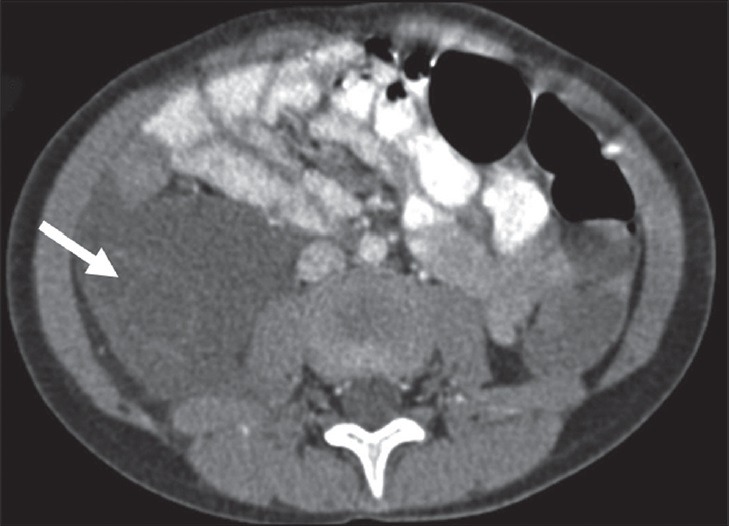
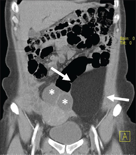
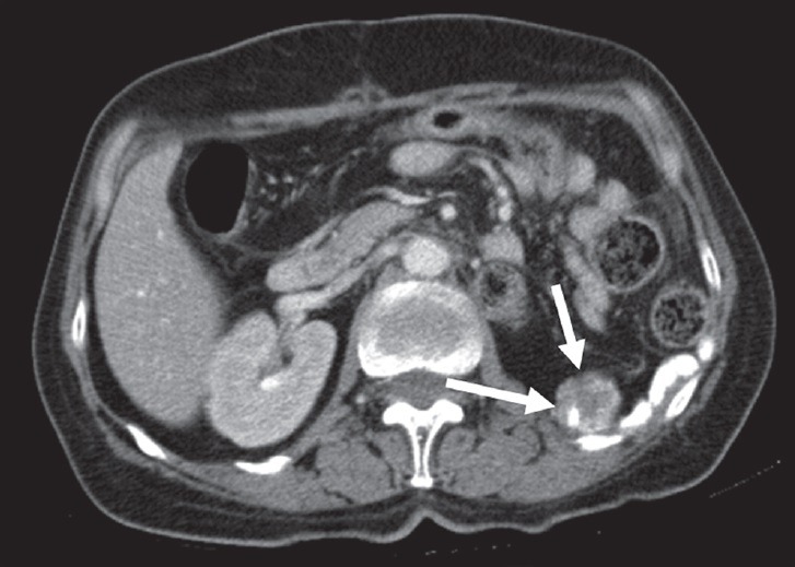
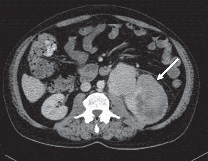
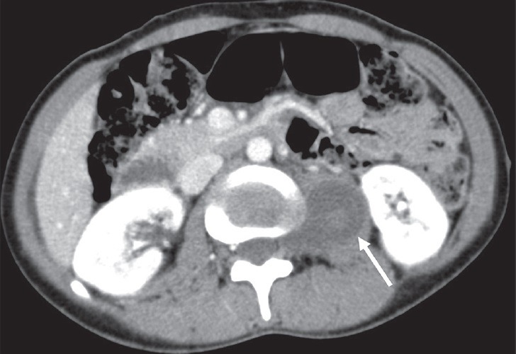
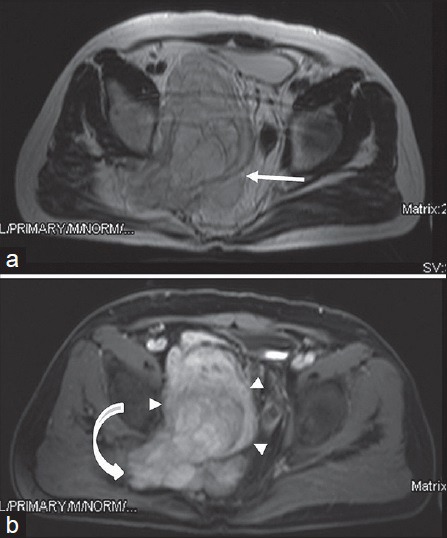
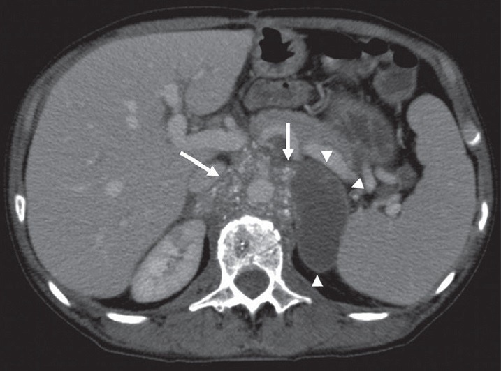
Basically malignant tumors in the retroperitoneal region arise from a heterogeneous group of tissues: mesodermal, neurogenic, germ cell, and lymphoid. Although rare, benign tumors and cystic masses can be also encountered in retroperitoneal space. Developments in computed tomography (CT) and magnetic resonance imaging (MRI) have contributed to both diagnosis and staging of the retroperitoneal tumors. High spatial resolution and superiority in calcification make CT indispensable; on the other hand, MRI has a better soft-tissue contrast resolution which is essential for the assessment of vascular invasion and tissue characterization. The aim of this article is to review the CT and MRI features of retroperitoneal tumors and their subsequent management.
基本上,腹膜后区域的恶性肿瘤起源于多种不同的组织:中胚层组织、神经源性组织、生殖细胞组织和淋巴组织。虽然少见,但腹膜后间隙也可出现良性肿瘤和囊性肿物。计算机断层扫描(CT)和磁共振成像(MRI)技术的发展对腹膜后肿瘤的诊断和分期均有帮助。CT具有高空间分辨率以及在显示钙化方面的优势,使其成为不可或缺的检查手段;另一方面,MRI具有更好的软组织对比分辨率,这对于评估血管侵犯和组织特征至关重要。本文旨在综述腹膜后肿瘤的CT和MRI特征及其后续治疗。